. 2024 Jun 3;65(6):7.
doi: 10.1167/iovs.65.6.7.

Radiation-Induced DNA Damage in Uveal Melanoma Is Influenced by Dose Delivery and Chromosome 3 Status

Affiliations

Radiation-Induced DNA Damage in Uveal Melanoma Is Influenced by Dose Delivery and Chromosome 3 Status

Aysegül Tura et al. Invest Ophthalmol Vis Sci. .

Abstract

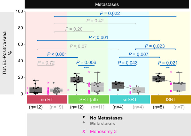
Purpose: The purpose of this study was to analyze the extent of DNA breaks in primary uveal melanoma (UM) with regard to radiotherapy dose delivery (single-dose versus fractionated) and monosomy 3 status.

Methods: A total of 54 patients with UM were included. Stereotactic radiotherapy (SRT) was performed in 23 patients, with 8 undergoing single-dose SRT (sdSRT) treatment and 15 receiving fractionated SRT (fSRT). DNA breaks in the enucleated or endoresected tumors were visualized by a TUNEL assay and quantified by measuring the TUNEL-positive area. Protein expression was analyzed by immunohistochemistry. Co-detection of chromosome 3 with proteins was performed by immuno-fluorescent in situ hybridization.

Results: The amount of DNA breaks in the total irradiated group was increased by 2.7-fold (P < 0.001) compared to non-irradiated tissue. Tumors treated with fSRT were affected more severely, showing 2.1-fold more DNA damage (P = 0.007) compared to the cases after single (high) dose irradiation (sdSRT). Monosomy 3 tumors showed less DNA breaks compared to disomy 3 samples (P = 0.004). The presence of metastases after radiotherapy correlated with monosomy 3 and less DNA breaks compared to patients with non-metastatic cancer in the combined group with fSRT and sdSRT (P < 0.05).

Conclusions: Fractionated irradiation led to more DNA damage than single-dose treatment in primary UM. As tumors with monosomy 3 showed less DNA breaks than those with disomy 3, this may indicate that they are less radiosensitive, which may influence the efficacy of irradiation.


Conflict of interest statement

Disclosure: A. Tura, Novartis Pharma GmbH, Germany (F); Y. Zhu, None; S. Vardanyan, None; M. Prasuhn, None; V. Kakkassery, None; J. Lüke, None; H. Merz, None; F. Paulsen, None; D. Rades, None; F. Cremers, None; K.-U. Bartz-Schmidt, None; S. Grisanti, None

Figures

Figure 1.
Figure 1.
Automated selection of the TUNEL-positive areas on the light microscopy images. The TUNEL reaction was visualized by using the horseradish peroxidase (HRP) green chromogen, whereas the nuclei were counterstained with nuclear fast red. Color deconvolution with user-defined settings was performed to separate the TUNEL signals (blue-green) from the nuclear staining and brown pigmentation with minimal overlap. The TUNEL panel was then processed for threshold adjustment for the automated selection of the positive regions. The sum of the TUNEL-positive areas was calculated and presented as the percentage of the total area that was analyzed in each tumor. Images were acquired at a magnification of 200 ×. Cells with a high versus low/negative TUNEL intensity are indicated by the black or purple arrows, respectively.
Figure 2.
Figure 2.
DNA damage in the combined cohort as demonstrated by the TUNEL-positive area. DNA damage was evaluated with regard to the (A) radiotherapy (RT) status, (B) frequency of stereotactic RT (SRT), (C) chromosome 3 copy number, (D) development of systemic metastases, and (E) metastasis-free survival. In (A to D), the ratio of the TUNEL-positive area to the total quantified area of each tumor was presented as percentage. The mean values were connected with the sloped lines. The red dots indicate the severe outliers. The P values were evaluated by the Mann-Whitney U test. fSRT, fractionated SRT; n, number; sdSRT, single-dose SRT. (E) Kaplan-Meier estimate of metastasis-free survival with respect to the DNA damage, which was classified based on the median TUNEL intensity (TUNEL-high: >median; TUNEL-low: <median). The x-axis indicates the time elapsed from the diagnosis of the primary tumor until the development of distant metastases or last follow-up. The y-axis demonstrates the percentage of patients that remained metastasis-free. Censored patients are indicated with the vertical ticks whereas the 95% confidence intervals are highlighted as background in the corresponding colors of each group. The number of patients at risk at each time point is presented underneath the curve. The P value was assessed by the log rank test.
Figure 3.
Figure 3.
DNA damage with regard to the chromosome 3 status in the tumors that received no radiotherapy (RT) versus stereotactic radiotherapy (SRT). The ratio of the TUNEL-positive area to the total quantified area of each tumor was stated as percentage. The mean values were connected with the sloped lines. The P values were determined by the Mann-Whitney U test. fSRT, fractionated SRT; n, number; sdSRT, single-dose SRT.
Figure 4.
Figure 4.
DNA damage with regard to the development of metastases in the tumors that received no radiotherapy (RT) versus stereotactic radiotherapy (SRT). The ratio of the TUNEL-positive area to the total quantified area of each tumor was indicated as percentage. The mean values were connected with the sloped lines. The tumors with monosomy 3 were indicated by the pinkx” symbols. The P values were assessed by the Mann-Whitney U test for the comparison of TUNEL-positive areas and by Fisher's Exact test for evaluating the ratios of monosomy 3 versus disomy 3 tumors with regard to the development of metastases in each group. **P < 0.005, ***P < 0.001 for the Fisher's Exact test. fSRT, fractionated SRT; n, number; sdSRT, single-dose SRT.

References

    1. Aronow ME, Topham AK, Singh AD.. Uveal melanoma: 5-year update on incidence, treatment, and survival (SEER 1973-2013). Ocul Oncol Pathol. 2018; 4(3): 145–151. - PMC - PubMed
    1. Krantz BA, Dave N, Komatsubara KM, Marr BP, Carvajal RD.. Uveal melanoma: epidemiology, etiology, and treatment of primary disease. Clin Ophthalmol. 2017; 11: 279–289. - PMC - PubMed
    1. Toro MD, Gozzo L, Tracia L, et al. .. New therapeutic perspectives in the treatment of uveal melanoma: a systematic review. Biomedicines. 2021; 9(10): 1311. - PMC - PubMed
    1. Blum ES, Yang J, Komatsubara KM, Carvajal RD.. Clinical management of uveal and conjunctival melanoma. Oncology (Williston Park). 2016; 30(1): 29–32, 34–43, 48. - PubMed
    1. Jager MJ, Shields CL, Cebulla CM, et al. .. Uveal melanoma. Nat Rev Dis Primers. 2020; 6(1): 24. - PubMed

MeSH terms