. 2024 Jun 4;24(24):7381-7388.
doi: 10.1021/acs.nanolett.4c01540. Online ahead of print.

Synergistic Biofilter Tube for Promoting Scarless Tendon Regeneration

Affiliations

Synergistic Biofilter Tube for Promoting Scarless Tendon Regeneration

Renhao Yang et al. Nano Lett. .

Abstract

Inspired by the imbalance between extrinsic and intrinsic tendon healing, this study fabricated a new biofilter scaffold with a hierarchical structure based on a melt electrowriting technique. The outer multilayered fibrous structure with connected porous characteristics provides a novel passageway for vascularization and isolates the penetration of scar fibers, which can be referred to as a biofilter process. In vitro experiments found that the porous architecture in the outer layer can effectively prevent cell infiltration, whereas the aligned fibers in the inner layer can promote cell recruitment and growth, as well as the expression of tendon-associated proteins in a simulated friction condition. It was shown in vivo that the biofilter process could promote tendon healing and reduce scar invasion. Herein, this novel strategy indicates great potential to design new biomaterials for balancing extrinsic and intrinsic healing and realizing scarless tendon healing.

Keywords: biofilter effects; biomedical melt electrowriting; hierarchical structure; scarless tendon healing.


Conflict of interest statement

The authors declare no competing financial interest.

Figures

Figure 1
Figure 1
Schematic diagram of the structure and the scarless tendon regeneration effect of the biofilter tube. (A) The structure of the biofilter tube for different directions. (B) The implantation and repair process of the biofilter tube in a rat tendon injury model. (C) The mechanism of the biofilter tube for scarless tendon regeneration, in which the neo fiber was aligned with the orientational scaffold fiber and the neovascularity penetrated in the scaffold and provided a local neovascularization environment.
Figure 2
Figure 2
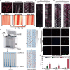
Anisotropic Janus tube scaffold characterization and local morphology. (A) The general morphology of the Janus tube and net patch scaffold. (B) Local architecture of the Janus scaffold. (C) Observation of the morphology of a Janus tube under an optical microscope. (D) SEM micrographs of the Janus tube from different sides. (E,F) SEM micrographs of the Janus tube from different sides after 5 days of cell culturing.
Figure 3
Figure 3
Influence of Janus tube on tenocyte behavior patterns and an in vitro shear simulation test. (A) Cytoskeleton staining in Janus tube and Net scaffold. (B) Cell distribution trend quantification on the scaffold surface. (C,D) Cell migration characteristics at different time points. (E) Schematic diagram of in vitro shear force simulation. (F) Expression of mechanics and tendon formation related proteins among different groups under shear force (bar = 100 μm). (G) Quantitative analysis of in vitro shear modeling (mean ± SD, Janus tube vs control, *** P < 0.001; Janus tube vs net scaffold, $ P < 0.05, $$ P < 0.01, $$$ P < 0.001; net scaffold vs control, # P < 0.05; n = 3).
Figure 4
Figure 4
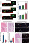
Evaluation repair performances of the Janus tube in vivo. (A) Application of different scaffolds for tendon repair. (B) Gross observation of peritendinous adhesion of injured tendon at 4 weeks. (C) Gross scores of peritendinous adhesion (mean ± SD; Janus tube vs control, *** P < 0.001; net scaffold vs control, ## P < 0.01; n = 3). (D) H&E staining images of different scaffolds. The dotted area showeds the adhesion region. (E,F) Histologic scores of tendon adhesion and tendon healing (mean ± SD, Janus tube vs control, *** P < 0.001; Janus tube vs net scaffold, $$$ P < 0.001; net scaffold vs control, # P < 0.05; n = 3). (G,H) Immunohistochemical staining and evaluation of Col I expression in peritendinous tissue at 4 weeks (mean ± SD; Janus tube vs control, *** P < 0.001; net scaffold vs control, ## P < 0.01; n = 3).
Figure 5
Figure 5
Assessment of Achilles tendon motor function and the histologic characteristics in the early phase of tendon healing. (A–C) Gait analysis of the repaired tendon at 4 weeks. (D) The printing area of the gait analysis (mean ± SD; Janus tube vs control, *** P < 0.001; Janus tube vs net scaffold, $$ P < 0.01; n = 3). (E) Comparison of maximum failure force between experimental groups. (F) H&E staining images of different scaffolds after 7 days. (bar = 100 μm) (mean ± SD; Janus tube vs control, *** P < 0.001; Janus tube vs net scaffold, $$ P < 0.01; net scaffold vs control, # P < 0.05; n = 3). (G) CD31 immunofluorescence staining for observing distribution of blood vessels in the tendon (bar = 100 μm). (H,I) Analysis of CD31 marked neovascularization patterns within and adjacent to the tendon sheath in different groups (Janus tube vs control, *P < 0.05; n = 3). (J) H&E staining and CD31 immunofluorescence staining of tendon cross-section in different groups (bar = 100 μm).

Similar articles

Cited by

References

    1. Nourissat G.; Berenbaum F.; Duprez D. Tendon injury: from biology to tendon repair. Nat. Rev. Rheumatol 2015, 11 (4), 223–233. 10.1038/nrrheum.2015.26. - DOI - PubMed
    1. Titan A. L.; Longaker M. T. A fine balance in tendon healing. Nat. Cell Biol. 2019, 21 (12), 1466–1467. 10.1038/s41556-019-0432-0. - DOI - PubMed
    1. Maffulli N.; Moller H. D.; Evans C. H. Tendon healing: can it be optimized?. Br J. Sports Med. 2002, 36 (5), 315–316. 10.1136/bjsm.36.5.315. - DOI - PMC - PubMed
    1. Deng B.; Xu P.; Zhang B.; Luo Q.; Song G. COX2 Enhances Neovascularization of Inflammatory Tenocytes Through the HIF-1alpha/VEGFA/PDGFB Pathway. Front Cell Dev Biol. 2021, 9, 670406.10.3389/fcell.2021.670406. - DOI - PMC - PubMed
    1. Li Y. W.; Ma C. H.; Huang H. K.; Lin K. J.; Wu C. H.; Tu Y. K. Use of Silicone Tubes as Antiadhesion Devices in a Modified Two-Stage Flexor Tendon Reconstruction in Zone II: A Retrospective Study. J. Hand Surg Am. 2023, 48 (5), 444–451. 10.1016/j.jhsa.2023.01.011. - DOI - PubMed