

Effects of an indole derivative on cell proliferation, transfection, and alternative splicing in production of lentiviral vectors by transient co-transfection
- PMID: 38833479
- PMCID: PMC11149887
- DOI: 10.1371/journal.pone.0297817
Effects of an indole derivative on cell proliferation, transfection, and alternative splicing in production of lentiviral vectors by transient co-transfection
Abstract
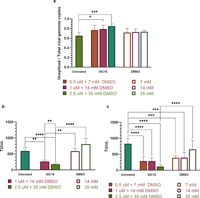
Lentiviral vectors derived from human immunodeficiency virus type I are widely used to deliver functional gene copies to mammalian cells for research and gene therapies. Post-transcriptional splicing of lentiviral vector transgene in transduced host and transfected producer cells presents barriers to widespread application of lentiviral vector-based therapies. The present study examined effects of indole derivative compound IDC16 on splicing of lentiviral vector transcripts in producer cells and corresponding yield of infectious lentiviral vectors. Indole IDC16 was shown previously to modify alternative splicing in human immunodeficiency virus type I. Human embryonic kidney 293T cells were transiently transfected by 3rd generation backbone and packaging plasmids using polyethyleneimine. Reverse transcription-quantitative polymerase chain reaction of the fraction of unspliced genomes in human embryonic kidney 293T cells increased up to 31% upon the indole's treatment at 2.5 uM. Corresponding yield of infectious lentiviral vectors decreased up to 4.5-fold in a cell transduction assay. Adjusting timing and duration of IDC16 treatment indicated that the indole's disruption of early stages of transfection and cell cycle had a greater effect on exponential time course of lentiviral vector production than its reduction of post-transcriptional splicing. Decrease in transfected human embryonic kidney 293T proliferation by IDC16 became significant at 10 uM. These findings indicated contributions by early-stage transfection, cell proliferation, and post-transcriptional splicing in transient transfection of human embryonic kidney 293T cells for lentiviral vector production.
Copyright: © 2024 Mier, Roper. This is an open access article distributed under the terms of the Creative Commons Attribution License, which permits unrestricted use, distribution, and reproduction in any medium, provided the original author and source are credited.
Conflict of interest statement
The authors have declared that no competing interests exist.
Figures


Similar articles
-
Production of lentiviral vectors for transducing cells from the central nervous system.J Vis Exp. 2012 May 24;(63):e4031. doi: 10.3791/4031. J Vis Exp. 2012. PMID: 22664962 Free PMC article.
-
The HIV-1 Rev protein enhances encapsidation of unspliced and spliced, RRE-containing lentiviral vector RNA.PLoS One. 2012;7(11):e48688. doi: 10.1371/journal.pone.0048688. Epub 2012 Nov 1. PLoS One. 2012. PMID: 23133650 Free PMC article.
-
Lentivirus production is influenced by SV40 large T-antigen and chromosomal integration of the vector in HEK293 cells.Hum Gene Ther. 2011 Oct;22(10):1269-79. doi: 10.1089/hum.2010.143. Epub 2011 Jun 13. Hum Gene Ther. 2011. PMID: 21554103
-
Large-scale production means for the manufacturing of lentiviral vectors.Curr Gene Ther. 2010 Dec;10(6):474-86. doi: 10.2174/156652310793797748. Curr Gene Ther. 2010. PMID: 21054245 Review.
-
Progress and Perspectives in the Development of Lentiviral Vector Producer Cells.Biotechnol J. 2021 Jan;16(1):e2000017. doi: 10.1002/biot.202000017. Epub 2020 Aug 2. Biotechnol J. 2021. PMID: 32686901 Review.
References
MeSH terms
Substances
LinkOut - more resources
Full Text Sources
