. 2024 Jun 4;20(6):e1012252.
doi: 10.1371/journal.ppat.1012252. eCollection 2024 Jun.

A specific innate immune response silences the virulence of Pseudomonas aeruginosa in a latent infection model in the Drosophila melanogaster host

Affiliations

A specific innate immune response silences the virulence of Pseudomonas aeruginosa in a latent infection model in the Drosophila melanogaster host

Jing Chen et al. PLoS Pathog. .

Abstract

Microbial pathogenicity often depends on the route of infection. For instance, P. aeruginosa or S. marcescens cause acute systemic infections when low numbers of bacteria are injected into D. melanogaster flies whereas flies succumb much slower to the continuous ingestion of these pathogens, even though both manage to escape from the gut compartment and reach the hemocoel. Here, we have developed a latent P. aeruginosa infection model by feeding flies on the bacteria for a short period. The bacteria stably colonize internal tissues yet hardly cause any damage since latently-infected flies live almost as long as noninfected control flies. The apparently dormant bacteria display particular characteristics in terms of bacterial colony morphology, composition of the outer cell wall, and motility. The virulence of these bacteria can however be reactivated upon wounding the host. We show that melanization but not the cellular or the systemic humoral response is the predominant host defense that establishes latency and may coerce the bacteria to a dormant state. In addition, the lasting activation of the melanization responses in latently-infected flies provides a degree of protection to the host against a secondary fungal infection. Latent infection by an ingested pathogen protects against a variety of homologous or heterologous systemic secondary infectious challenges, a situation previously described for the endosymbiotic Wolbachia bacteria, a guard against viral infections.


Conflict of interest statement

The authors have declared that no competing interests exist.

Figures

Fig 1
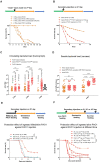
Fig 1. P. aeruginosa that have escaped from the gut lumen into the body cavity lose pathogenicity and may become dormant in Drosophila.
(A) Scheme of P. aeruginosa latent infection model in Drosophila. Flies were fed with 10 OD600 PAO1 suspended in 100mM sucrose solution with 10 percent Brain-Heart Infusion broth for 2 days and then PAO1 in gut lumen were killed by feeding sucrose solution containing 100μg/mL gentamicin for another 4 days, leaving alive only the bacteria that had crossed the digestive tract. Then flies were fed sucrose solution until death. Flies were put at 18°C throughout the infection process. (B) Survival curves after latent infection. (C) Bacterial titer from collected hemolymph. Each dot indicates the bacterial number collected from a single fly. N, the number of flies, is indicated above the data for each time point; the first number corresponds to the number of flies for which no bacteria were retrieved (0) and the second number corresponds to the number of flies for which bacteria were retrieved. (D) Time course of bacterial titer in whole flies (filled circles) and carcass (empty squares). Each dot indicates the bacterial number retrieved from a single fly or a single carcass. The numbers above each column indicate the median value of the bacterial titer in each column. (E-F) P. aeruginosa visualization in fly tissue. (E) P. aeruginosa PAO1 was visualized by HOECHST staining. (F) P. aeruginosa PA14 was stained with an antibody raised against it. (G) Activation of the IMD pathway upon ingested P. aeruginosa infection as monitored by RTqPCR of Diptericin transcripts normalized to rpl49 transcript levels of each sample. (B-G). All experiments here were performed three times independently, and data were pooled together (B, C, D, G). The bar indicates the median in each column (C, D and G). Statistical analysis was done by Logrank (Mantel-Cox test) in (B) or by Mann-Whitney test in (D and G).
Fig 2
Fig 2. P. aeruginosa bacteria colonizing tissues in the body cavity exhibit distinct traits from injected ones.
(A-C) Morphology of P. aeruginosa in the latent infection model (A), in the injection model (B), and cultured in BHB (C). Hemolymph was collected from flies with injected or ingested PAO1 respectively and the bacteria were observed by transmission electron microscopy. Scale bars are 500 nm. (D) LPS O5 antigen staining (green) of RFP-expressing bacteria retrieved from hemolymph (top panels) or from tissues (bottom panels). Note the absence of staining in bacteria colonizing the tissues in the latent infection model. (E) Colonies of bacteria retrieved from infected flies (at Day6 for latently-infected flies and at Day2 for injected flies), as indicated, forming on Congo RED agar plates. In vitro cultures of PAO1 were in BHB medium. (F) Motility assay of bacteria retrieved from infected flies, as indicated, on low percentage agar plates. (G and G’) Sensitivity of bacteria in vivo to 4.6 nL-injected tobramycin (TOB: 16mg/mL) or levofloxacin (8mg/mL) measured by assessing the bacterial load of single whole flies at the indicated times after antibiotics injection in the latent (G) or injection (G’) infection model. (H) Scheme of pathogenicity experiment for PAO1 retrieved from latently-infected flies. Hemolymph was collected from flies with ingested PAO1 at Day 2 post feeding and injected into naive flies in couples. (H’) Survival curves of naïve flies to injected cultured P. aeruginosa (red curve) or P. aeruginosa retrieved from latently-infected flies (orange curve). All experiments were performed three times independently; data were pooled (G, G’, and H’). The bar indicates the median in each column (G and G’). Statistical analysis was done using the Kruskal-Wallis test with Dunn’s post-hoc test (G and G’), or by Logrank (Mantel-Cox test) in (H’).
Fig 3
Fig 3. Melanization is the preponderant host defense that initiates dormancy in the bacteria that have escaped from the digestive tract.
(A) Survival curves for key and eater deficient flies. (B) Bacterial titer in the hemolymph collected from key and eater deficient flies. Each dot corresponds to the bacterial load of a single fly. (C) Survival curves of melanization-deficient flies: Hayan, ΔPPO1-ΔPPO2, and Sp7. (D) Bacterial titer in the hemolymph collected from wild-type flies or mutant for phenol oxidase genes. (E) Bacterial titer of single fly carcasses of prophenoloxidase-deficient flies. (F) PPO cleavage in different infection models as revealed by Western blot analysis using a pan-PO reactive antibody. All experiments here were performed three times independently and data were pooled together (A-E). The bar indicates the median in each column (B, D and E). Statistical analysis was done using Logrank (Mantel-Cox test) in (A and C), and Mann-Whitney test in (B), Kruskal-Wallis with Dunn’s post-hoc test in (D and E).
Fig 4
Fig 4. The immune responses elicited by ingested P. aeruginosa provide protection against a secondary P. aeruginosa injection.
(A) Injury on flies at day 2 activates full virulence of ingested P. aeruginosa, as monitored in survival experiments. Injuries were performed either by pricking with a sharp needle, or by collecting hemolymph or injecting PBS with a micro-pipette. (B) Survival curves of flies submitted to a secondary infection by injecting cultured RFP-labeled PAO1 to flies that had either ingested GFP-labeled PAO1 or a control sucrose solution. (C-D) Time course of bacterial burden of flies infected as in (B); the bacteria were collected either from the hemolymph or from carcasses of single flies. (E) The ingestion of heat-killed P. aeruginosa elicits a mild protection against injected PAO1 as compared to that induced by the ingestion of live P. aeruginosa, as monitored in survival experiments. (F) The protection afforded by the ingestion of live PAO1 becomes stronger as the secondary injection challenge is performed later and later. The protection progressively increases when the secondary injection is performed at day 6 and then day 10, as compared to the initial day 2 challenge when bacteria have not been cleared from the gut lumen. All experiments here were performed three times independently (4A excepted, one time) and data were pooled together (A-F). The bar indicates the median in each column (C and D). Statistical analysis was done using the Logrank test (Mantel-Cox test) in (A, B, E, and F) and using the Kruskal-Wallis test with Dunn’s post-hoc test in (C and D).
Fig 5
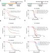
Fig 5. The silent colonization by PAO1 in the tissues confers to the latently-infected flies an enhanced protection against secondary bacterial or fungal infections that is likely mediated by melanization.
(A-C) Survival curves of secondary infection by the Gram-negative bacterium Serratia marcescens (A), the Gram-positive Enterococcus faecalis (B), and the fungal entomopathogen Metarhizium robertsii in PAO1-latently infected flies as compared to the same infection in naïve flies fed with sucrose solution (C). (D-F) Impact of mutations impairing different enzymes involved in melanization on the enhanced defense conferred by the prior ingestion of PAO1: ΔPPO1-ΔPPO2 (D), Hayan (E), and Sp7 (F). Because these mutants are susceptible to the ingestion of PAO1, the virulence in immunodeficient flies of PAO1 bacteria colonizing the tissues was mitigated by the oral administration of 100μg/mL Levofloxacin for four days, following the ingestion of PAO1 for two days. The M. robertsii fungus is insensitive to the action of the antibiotics. All experiments here were performed three times independently and data were pooled together. Statistical analysis was done using the Logrank test (Mantel-Cox test) in (A-F).

Similar articles

Cited by

References

    1. Thi MTT, Wibowo D, Rehm BHA. Pseudomonas aeruginosa Biofilms. International journal of molecular sciences. 2020;21(22):8671. doi: 10.3390/ijms21228671 - DOI - PMC - PubMed
    1. Fisher RA, Gollan B, Helaine S. Persistent bacterial infections and persister cells. Nat Rev Microbiol. 2017;15(8):453–64. doi: 10.1038/nrmicro.2017.42 - DOI - PubMed
    1. Pang Z, Raudonis R, Glick BR, Lin TJ, Cheng Z. Antibiotic resistance in Pseudomonas aeruginosa: mechanisms and alternative therapeutic strategies. Biotechnology advances. 2019;37(1):177–92. doi: 10.1016/j.biotechadv.2018.11.013 - DOI - PubMed
    1. Boman HG, Nilsson I, Rasmuson B. Inducible antibacterial defence system in Drosophila. Nature. 1972;237(5352):232–5. doi: 10.1038/237232a0 - DOI - PubMed
    1. Limmer S, Quintin J, Hetru C, Ferrandon D. Virulence on the fly: Drosophila melanogaster as a model genetic organism to decipher host-pathogen interactions. Current Drug Targets. 2011;12(7):978–99. doi: 10.2174/138945011795677818 - DOI - PubMed

MeSH terms

LinkOut - more resources