

Shared Mechanisms Drive Ocular Following and Motion Perception
- PMID: 38834301
- PMCID: PMC11208981
- DOI: 10.1523/ENEURO.0204-24.2024
Shared Mechanisms Drive Ocular Following and Motion Perception
Abstract
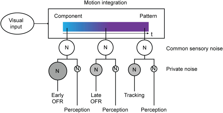
How features of complex visual patterns are combined to drive perception and eye movements is not well understood. Here we simultaneously assessed human observers' perceptual direction estimates and ocular following responses (OFR) evoked by moving plaids made from two summed gratings with varying contrast ratios. When the gratings were of equal contrast, observers' eye movements and perceptual reports followed the motion of the plaid pattern. However, when the contrasts were unequal, eye movements and reports during early phases of the OFR were biased toward the direction of the high-contrast grating component; during later phases, both responses followed the plaid pattern direction. The shift from component- to pattern-driven behavior resembles the shift in tuning seen under similar conditions in neuronal responses recorded from monkey MT. Moreover, for some conditions, pattern tracking and perceptual reports were correlated on a trial-by-trial basis. The OFR may therefore provide a precise behavioral readout of the dynamics of neural motion integration for complex visual patterns.
Keywords: eye movements; ocular following; pattern motion; perception–action.
Copyright © 2024 Kreyenmeier et al.
Conflict of interest statement
The authors declare no competing financial interests.
Figures


Update of
-
Shared mechanisms drive ocular following and motion perception.bioRxiv [Preprint]. 2023 Oct 2:2023.10.02.560543. doi: 10.1101/2023.10.02.560543. bioRxiv. 2023. Update in: eNeuro. 2024 Jun 20;11(6):ENEURO.0204-24.2024. doi: 10.1523/ENEURO.0204-24.2024. PMID: 37873151 Free PMC article. Updated. Preprint.
Similar articles
-
Shared mechanisms drive ocular following and motion perception.bioRxiv [Preprint]. 2023 Oct 2:2023.10.02.560543. doi: 10.1101/2023.10.02.560543. bioRxiv. 2023. Update in: eNeuro. 2024 Jun 20;11(6):ENEURO.0204-24.2024. doi: 10.1523/ENEURO.0204-24.2024. PMID: 37873151 Free PMC article. Updated. Preprint.
-
Perceptual, oculomotor, and neural responses to moving color plaids.Perception. 1998;27(6):681-709. doi: 10.1068/p270681. Perception. 1998. PMID: 10197187
-
Tracking without perceiving: a dissociation between eye movements and motion perception.Psychol Sci. 2011 Feb;22(2):216-25. doi: 10.1177/0956797610394659. Epub 2010 Dec 28. Psychol Sci. 2011. PMID: 21189353 Free PMC article.
-
Temporal dynamics of 2D motion integration for ocular following in macaque monkeys.J Neurophysiol. 2010 Mar;103(3):1275-82. doi: 10.1152/jn.01061.2009. Epub 2009 Dec 23. J Neurophysiol. 2010. PMID: 20032230
-
Responses of neurons in macaque MT to unikinetic plaids.J Neurophysiol. 2019 Nov 1;122(5):1937-1945. doi: 10.1152/jn.00486.2019. Epub 2019 Sep 11. J Neurophysiol. 2019. PMID: 31509468 Free PMC article.
Cited by
-
Inability to pursue nonrigid motion produces instability of spatial perception.Sci Adv. 2024 Nov 8;10(45):eadp6204. doi: 10.1126/sciadv.adp6204. Epub 2024 Nov 6. Sci Adv. 2024. PMID: 39504371 Free PMC article.
-
Poor fixation stability does not account for motion perception deficits in amblyopia.Sci Rep. 2025 Jan 25;15(1):3183. doi: 10.1038/s41598-024-83624-9. Sci Rep. 2025. PMID: 39863630 Free PMC article.
-
Probabilistically constrained vector summation of motion direction in the mouse superior colliculus.Curr Biol. 2025 Feb 24;35(4):723-733.e3. doi: 10.1016/j.cub.2024.12.029. Epub 2025 Jan 21. Curr Biol. 2025. PMID: 39842438
References
MeSH terms
Grants and funding
LinkOut - more resources
Full Text Sources