

Computed tomographic evaluation of portal vein indices in cats with the extrahepatic portosystemic shunts
- PMID: 38834507
- PMCID: PMC11156598
- DOI: 10.4142/jvs.24038
Computed tomographic evaluation of portal vein indices in cats with the extrahepatic portosystemic shunts
Abstract
Importance: The portal vein to aorta (PV/Ao) ratio is used to assess the clinical significance of extrahepatic portosystemic shunt (EHPSS). Previous studies using computed tomography (CT) were conducted in dogs but not in cats.
Objective: This study aimed to establish normal reference values for PV indices (PV/Ao ratio and PV diameter) in cats and determine the usefulness of these for predicting symptomatic EHPSS.
Methods: This study included 95 dogs and 114 cats that underwent abdominal CT. The canine normal (CN) group included dogs without EHPSS. The cats were classified into feline normal (FN, 88/114), feline asymptomatic (FA, 16/114), and feline symptomatic (FS, 10/114) groups. The PV and Ao diameters were measured in axial cross-sections.
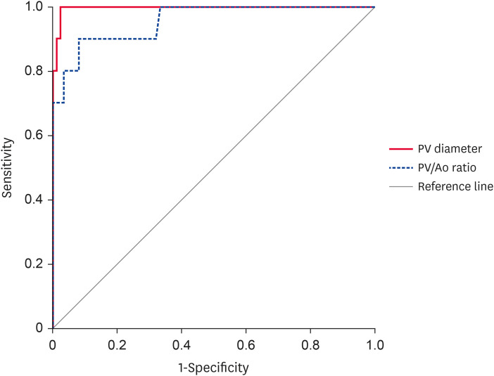
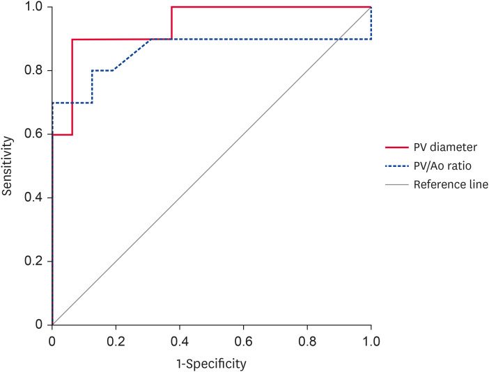
Results: The group FN had a higher PV/Ao ratio than the group CN (p < 0.001). Within the feline groups, the PV indices were in the order FN > FA > FS (both p < 0.001). The mean PV diameter and PV/Ao ratio for group FN were 5.23 ± 0.77 mm and 1.46 ± 0.19, respectively. The cutoff values between groups FN and FS were 4.115 mm for PV diameter (sensitivity, 100%; specificity, 97.7%) and 1.170 for PV/Ao ratio (90%, 92.1%). The cutoff values between group FA and FS were 3.835 mm (90%, 93.8%) and 1.010 (70%, 100%), respectively.
Conclusions and relevance: The results demonstrated significant differences in PV indices between dogs and cats. In cats, the PV/Ao ratio demonstrated high diagnostic performance for symptomatic EHPSS. The PV diameter also performed well, in contrast to dogs.
Keywords: CT; PSS; PV diameter; PV/Ao ratio; feline.
© 2024 The Korean Society of Veterinary Science.
Conflict of interest statement
The authors declare no conflicts of interest.
Figures
Similar articles
-
Ultrasonographic diagnosis of portosystemic shunting in dogs and cats.Vet Radiol Ultrasound. 2004 Sep-Oct;45(5):424-37. doi: 10.1111/j.1740-8261.2004.04076.x. Vet Radiol Ultrasound. 2004. PMID: 15487568
-
Portal Vein/Aorta Ratio in Dogs with Acquired Portosystemic Collaterals.J Vet Intern Med. 2017 Sep;31(5):1382-1387. doi: 10.1111/jvim.14802. Epub 2017 Aug 14. J Vet Intern Med. 2017. PMID: 28804949 Free PMC article.
-
Anatomical classification of feline congenital extrahepatic portosystemic shunts based on CT angiography: A SVSTS and VIRIES multi-institutional study in 231 cats.Vet Radiol Ultrasound. 2024 Jul;65(4):359-368. doi: 10.1111/vru.13363. Epub 2024 Apr 10. Vet Radiol Ultrasound. 2024. PMID: 38597362
-
Ultrasonography of portosystemic shunts in dogs and cats.Vet Clin North Am Small Anim Pract. 1998 Jul;28(4):725-53. doi: 10.1016/s0195-5616(98)50076-9. Vet Clin North Am Small Anim Pract. 1998. PMID: 9698613 Review.
-
Diagnostic imaging of congenital porto-systemic shunts in dogs and cats: a review.Vet J. 2003 Jul;166(1):7-18. doi: 10.1016/s1090-0233(02)00304-0. Vet J. 2003. PMID: 12788013 Review.
References
-
- Watson PJ, Herrtage ME. Medical management of congenital portosystemic shunts in 27 dogs--a retrospective study. J Small Anim Pract. 1998;39(2):62–68. - PubMed
-
- Winkler JT, Bohling MW, Tillson DM, Wright JC, Ballagas AJ. Portosystemic shunts: diagnosis, prognosis, and treatment of 64 cases (1993–2001) J Am Anim Hosp Assoc. 2003;39(2):169–185. - PubMed
-
- Payne JT, Martin RA, Constantinescu GM. The anatomy and embryology of portosystemic shunts in dogs and cats. Semin Vet Med Surg Small Anim. 1990;5(2):76–82. - PubMed
-
- Cabassu J, Seim HB, 3rd, MacPhail CM, Monnet E. Outcomes of cats undergoing surgical attenuation of congenital extrahepatic portosystemic shunts through cellophane banding: 9 cases (2000–2007) J Am Vet Med Assoc. 2011;238(1):89–93. - PubMed
-
- Hunt GB. Effect of breed on anatomy of portosystemic shunts resulting from congenital diseases in dogs and cats: a review of 242 cases. Aust Vet J. 2004;82(12):746–749. - PubMed
MeSH terms
LinkOut - more resources
Full Text Sources
Medical
Miscellaneous
