

Extracellular fluid excess linked to reduced choroidal vascularity index in patients with chronic kidney disease
- PMID: 38834727
- PMCID: PMC11150457
- DOI: 10.1038/s41598-024-63444-7
Extracellular fluid excess linked to reduced choroidal vascularity index in patients with chronic kidney disease
Abstract
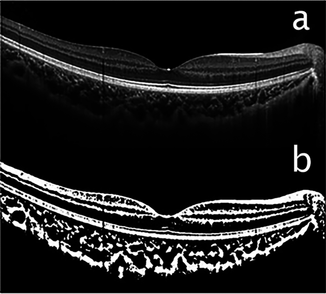
Extracellular fluid (ECF) excess is common in patients with chronic kidney disease (CKD). This study (involving 284 patients with CKD) explored the association between choroidal vascularity index (CVI) and ECF excess. We categorised patients into three groups based on extracellular water/total body water: normal, mildly overhydrated, and severely overhydrated. The more severe ECF status was associated with a lower CVI after adjustment (B = - 0.902, p = 0.001). In non-diabetic patients, both vascular luminal (LA, p < 0.001) and stromal areas (SA, p = 0.003) were significantly reduced in patients with severe ECF excess compared to others, whereas diabetic patients showed no significant differences in LA (p = 0.96) and SA (p = 0.86) based on ECF excess status. These findings suggest that ECF status may influence CVI in patients with CKD, underscoring the need for further research to clarify its direct impact on choroidal changes.
© 2024. The Author(s).
Conflict of interest statement
The authors declare no competing interests.
Figures
Similar articles
-
Choroidal vascularity index as a predictor for the development of retinopathy in diabetic patients.J Endocrinol Invest. 2024 May;47(5):1175-1180. doi: 10.1007/s40618-023-02236-8. Epub 2023 Nov 22. J Endocrinol Invest. 2024. PMID: 37993663
-
[Features of structural and microvascular changes of the choroid in angioretinopathy of various etiologies].Vestn Oftalmol. 2022;138(2):47-56. doi: 10.17116/oftalma202213802147. Vestn Oftalmol. 2022. PMID: 35488562 Russian.
-
Vascular changes of the choroid and their correlations with visual acuity in diabetic retinopathy.Front Endocrinol (Lausanne). 2024 Feb 23;15:1327325. doi: 10.3389/fendo.2024.1327325. eCollection 2024. Front Endocrinol (Lausanne). 2024. PMID: 38464970 Free PMC article.
-
Choroidal vascularity index: a step towards software as a medical device.Br J Ophthalmol. 2022 Feb;106(2):149-155. doi: 10.1136/bjophthalmol-2021-318782. Epub 2021 Jan 29. Br J Ophthalmol. 2022. PMID: 33514528 Review.
-
Choroidal Perfusion Changes After Vitrectomy for Myopic Traction Maculopathy.Semin Ophthalmol. 2024 May;39(4):261-270. doi: 10.1080/08820538.2023.2283029. Epub 2023 Nov 21. Semin Ophthalmol. 2024. PMID: 37990380 Review.
References
MeSH terms
Grants and funding
- 6-2023-0160/Faculty research grant from Yonsei University College of Medicine
- 6-2023-0160/Faculty research grant from Yonsei University College of Medicine
- 6-2023-0160/Faculty research grant from Yonsei University College of Medicine
- 6-2023-0160/Faculty research grant from Yonsei University College of Medicine
- 6-2023-0160/Faculty research grant from Yonsei University College of Medicine
LinkOut - more resources
Full Text Sources
Medical
