. 2024 Jul;131(2):258-270.
doi: 10.1038/s41416-024-02724-5. Epub 2024 Jun 4.

CD57 defines a novel cancer stem cell that drive invasion of diffuse pediatric-type high grade gliomas

Affiliations

CD57 defines a novel cancer stem cell that drive invasion of diffuse pediatric-type high grade gliomas

Lin Qi et al. Br J Cancer. 2024 Jul.

Abstract

Background: Diffuse invasion remains a primary cause of treatment failure in pediatric high-grade glioma (pHGG). Identifying cellular driver(s) of pHGG invasion is needed for anti-invasion therapies.

Methods: Ten highly invasive patient-derived orthotopic xenograft (PDOX) models of pHGG were subjected to isolation of matching pairs of invasive (HGGINV) and tumor core (HGGTC) cells.

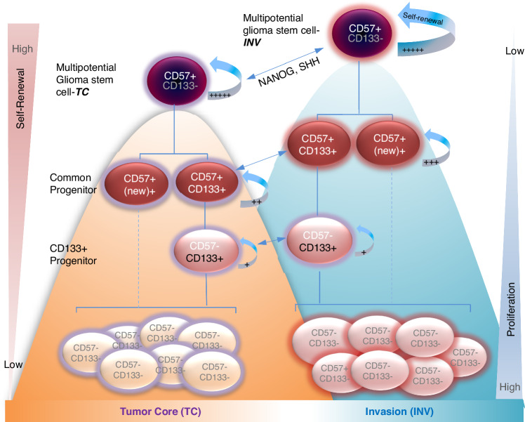
Results: pHGGINV cells were intrinsically more invasive than their matching pHGGTC cells. CSC profiling revealed co-positivity of CD133 and CD57 and identified CD57+CD133- cells as the most abundant CSCs in the invasive front. In addition to discovering a new order of self-renewal capacities, i.e., CD57+CD133- > CD57+CD133+ > CD57-CD133+ > CD57-CD133- cells, we showed that CSC hierarchy was impacted by their spatial locations, and the highest self-renewal capacities were found in CD57+CD133- cells in the HGGINV front (HGGINV/CD57+CD133- cells) mediated by NANOG and SHH over-expression. Direct implantation of CD57+ (CD57+/CD133- and CD57+/CD133+) cells into mouse brains reconstituted diffusely invasion, while depleting CD57+ cells (i.e., CD57-CD133+) abrogated pHGG invasion.

Conclusion: We revealed significantly increased invasive capacities in HGGINV cells, confirmed CD57 as a novel glioma stem cell marker, identified CD57+CD133- and CD57+CD133+ cells as a new cellular driver of pHGG invasion and suggested a new dual-mode hierarchy of HGG stem cells.


Conflict of interest statement

The authors declare no competing interests.

Figures

Fig. 1
Fig. 1. PDOX HGG tumors were highly invasive in vivo.
a Detection and quantitation of HGG invasion through IHC staining using human-specific antibodies against vimentin (VMT) or mitochondria (MT) (arrow). Representative images showed invasion modes through single cells, along blood vessels (perivascular invasion), along neural fibers, and spread through cerebral spinal fluid (CSF) (upper panel). The distances of invasion between the most distant (or deepest into the normal brains) invasive cells the border line of tumor core (black dotted line) in four PDOX models were measured and graphed (lower panel). Scale bar = 100 µM. b Representative gross appearance of HGG PDOX model showing intra-cerebral xenograft formation (arrow). c Strategies of isolating matching pairs of HGGINV and HGGTC cells. Whole mouse brains sliced at 1 mm thickness into 10–12 slices. The tumor core was dissected from “normal” mouse brains under stereotactic microscope. “Normal” mouse brain tissues (containing HGGINV cells) and tumor mass (containing HGGTC cells) were placed in cold (4 °C) growth medium in separate Petri dishes and dissociated into single cell suspension using Gentle Dissociator (Miltenyi). d FACS decontamination of mouse cells. Cell suspensions were incubated with FITC-conjugated monoclonal antibodies against human HLA-ABC and APC-conjugated monoclonal antibodies against mouse major histocompatibility antigen. Mouse cells (APC-positive and FITC-negative) were gated out together with the dead cells (propidium iodine high).
Fig. 2
Fig. 2. HGGINV cells possess stronger invasive capacity than the matching HGGTC cells.
Serial passaging of the purified 1406HGGTC (TC1 ~ TC3) (a) and 1406HGGINV (INV1-INV3) (b) cells in vivo in mouse brains reveal gradually decreased invasion in HGGTC cells and significantly enhanced invasion of HGGINV cells as detected by IHC staining using human-specific antibodies against VMT (arrow) (upper panel). Scale bar = 100 µM. c The distances between the most distant cells in the invasive front and the border line of tumor core (black dotted line) were measured and graphed (lower panel) (P < 0.01). d Changes of animal survival times from 3 PDOX models during serial transplantation of HGGTC and HGGINV cells through log-rank analysis. All the mice received intra-cerebral implantation of identical number of tumor cells (1 × 105 in 2 µL) from 3 PDOX models IC-1406HGG (1406HGG), IC-2305HGG (2305HGG) and IC-3752HGG (3752HGG).
Fig. 3
Fig. 3. HGG invasive cells have stronger neurosphere forming capacity than the matching HGGTC cells in vitro.
a Comparison of neurosphere formation efficiencies between HGGTC and HGGINV cells of 6 PDOX models. The HGG cells were cultured in CSC medium for up to 14 days in triplicates through serial dilution in 96-well plates. Date presented as mean ± SD. HGGHGGHGGHGGHGGHGGHGG. b Expression of self-renewal genes between HGGINV and HGGTC cells derived from the 6 PDOX models as detected by q-RT-PCR (**P < 0.01).
Fig. 4
Fig. 4. CD57 as new marker HGG CSCs.
a FCM profiling of putative CSC markers in 8 PDOX tumor cells of HGG. b Quantitative analysis of CD57 and CD133 expression in patient HGG tumors through FCM (left panel). Tumor cells with mono- and dual-positive CD57 and CD133 were graphed from 13 patient HGGs (right panel). Each sample was analyzed once. c High level expression of CD57 and expression of CD133 in 15 PDOX models of HGG as detected by FCM. d CD57+ cells were enriched in the invasive front (HGGINV cells), while CD133+ cells were mostly in the tumor core (HGGTC groups) in four HGG models (**P < 0.05).
Fig. 5
Fig. 5. Elevated invasion and self-renewal capacities of CD57+ tumor cells in the HGGINV fraction in vitro.
HGGTC and HGGINV cells PDOX models were further fractionated into CD57+ and/or CD133+ (CD133-CD57+, CD133+CD57+, CD133+CD57 and CD133CD57) via FACS for quantitative and functional assays. a Evaluation of in vitro self-renewal capacity of mono- and dual-positive CD57 HGG cells as compared with CD133 tumor cells. High-content image analysis was performed 14 days post tumor cell incubation (upper panel) to quantitate the neurosphere size and number of the whole wells of 96-well plates. Three models were included. b HGGINV/CD57+CD133- cells exhibited the strongest self-renewal capacity in vitro in cells derived from all four HGG models (1128HGG, 2305HGG, 1406HGG and 3752HGG) (P < 0.01). c HGGINV/ CD57+ cells expressed high levels of SHH and NANOG genes as detected by q-RT-PCR. Data were presented as the ratios of HGGINV vs HGGTC (Mean ± SD) from three replicates.
Fig. 6
Fig. 6. CD57+ cells as cellular driver of HGG invasion.
a Tumorigenicity of the fractionated CD57 and/or CD133 positive cells derived from HGGINV and HGGTC cells. Each mouse received 100 to 1000 purified tumor cells. b CD57+ and/or CD133+ cells in the invasive front (HGGINVcells) exhibited stronger invasion capacity in vitro. Tumor cells from three HGG models (1406HGG, 2305HGG and 3752HGG) were seeded in triplicate using CytoSelect 24/96-Well Cell invasion kit (upper panel). Tumor cells that migrated through the membrane (arrow) were quantitatively compared between tumor core (HGGTC) and their matching invasive (HGGINV) cells and graphed as Mean ± SD (**P < 0.01). Dual-negative (CD57CD133) cells were included as control. c In vivo evaluation of the invasive capacity of fractionated CD57+ and/or CD133+ glioma cells isolated from the invasive front (INV) and compared with those from the tumor core (TC). Human tumor cells were identified by IHC staining using human-specific antibodies against vimentin (VMT) and mitochondria (MT) (Scale bar = 100 µM). The depth of invasion was measured from the most distant cells of the invasive front to the border line of tumor core (black line) and graphed. Slides incubated without the primary antibodies were uses as control. Data presented as Mean ± SD from 3 sections. (*P < 0.01). d Immunohistochemical staining of C57, GFAP, Ki67, NeuN and Nestin in xenografts derived from spatial (INV v.s TC) related CD57+ and/or CD133+ pHGG cells.
Fig. 7
Fig. 7. Spatial related dual-mode CSC hierarchy of pHGG.
Situated on the top are CD57+CD133 cells that represent the multipotential glioma stem cells, followed by CD57+CD133+ cells as common progenitor cells that give rise to CD57CD133+ cells that further differentiate to non-stem CD57CD133 tumor cells. Due to the spatial/location differences, those who migrated into normal brain cells, particularly the HGGINV/CD57+CD133 cells, gained extra self-renewal capacity than the HGGTC/CD57+CD133+ cells, possibly mediated by NANOG and SHH. The differences of elevated self-renewal capacity of common and CD133+ progenitor cells between HGGINV and HGGTC cells progressively decrease following the formation and expansion of invasive satellite tumor mass; such dynamic changes are indicated by the bidirectional arrows. Cell proliferation capacity is reversely correlated with stemness (e.g., self-renewal capacity); with the non-stem dual-negative (CD57CD133) cells exhibiting the highest proliferative capacity. In addition to CD133, there may be new/unknown common progenitors expressing additional marker(s).

Similar articles

References

    1. Louis DN, Perry A, Wesseling P, Brat DJ, Cree IA, Figarella-Branger D, et al. The 2021 WHO classification of tumors of the central nervous system: a summary. Neuro Oncol. 2021;23:1231–51. 10.1093/neuonc/noab106 - DOI - PMC - PubMed
    1. Baxter PA, Su JM, Onar-Thomas A, Billups CA, Li XN, Poussaint TY, et al. A phase I/II study of veliparib (ABT-888) with radiation and temozolomide in newly diagnosed diffuse pontine glioma: a Pediatric Brain Tumor Consortium study. Neuro Oncol. 2020;22:875–85. 10.1093/neuonc/noaa016 - DOI - PMC - PubMed
    1. Tsoli M, Shen H, Mayoh C, Franshaw L, Ehteda A, Upton D, et al. International experience in the development of patient-derived xenograft models of diffuse intrinsic pontine glioma. J Neurooncol. 2019;141:253–63. 10.1007/s11060-018-03038-2 - DOI - PubMed
    1. Stupp R, Mason WP, van den Bent MJ, Weller M, Fisher B, Taphoorn MJ, et al. Radiotherapy plus concomitant and adjuvant temozolomide for glioblastoma. N. Engl J Med. 2005;352:987–96. 10.1056/NEJMoa043330 - DOI - PubMed
    1. Stupp R, Hegi ME, Mason WP, van den Bent MJ, Taphoorn MJ, Janzer RC, et al. Effects of radiotherapy with concomitant and adjuvant temozolomide versus radiotherapy alone on survival in glioblastoma in a randomised phase III study: 5-year analysis of the EORTC-NCIC trial. Lancet Oncol. 2009;10:459–66. 10.1016/S1470-2045(09)70025-7 - DOI - PubMed