. 2024 Aug;34(8):545-555.
doi: 10.1038/s41422-024-00981-w. Epub 2024 Jun 4.

Molecular and structural basis of an ATPase-nuclease dual-enzyme anti-phage defense complex

Affiliations

Molecular and structural basis of an ATPase-nuclease dual-enzyme anti-phage defense complex

Qiyin An et al. Cell Res. 2024 Aug.

Abstract

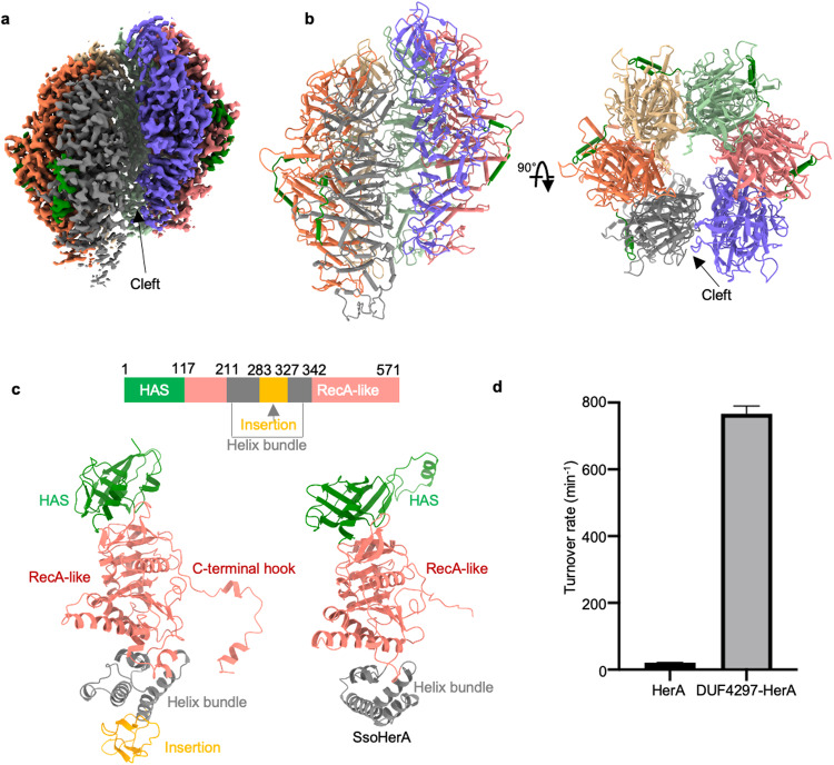
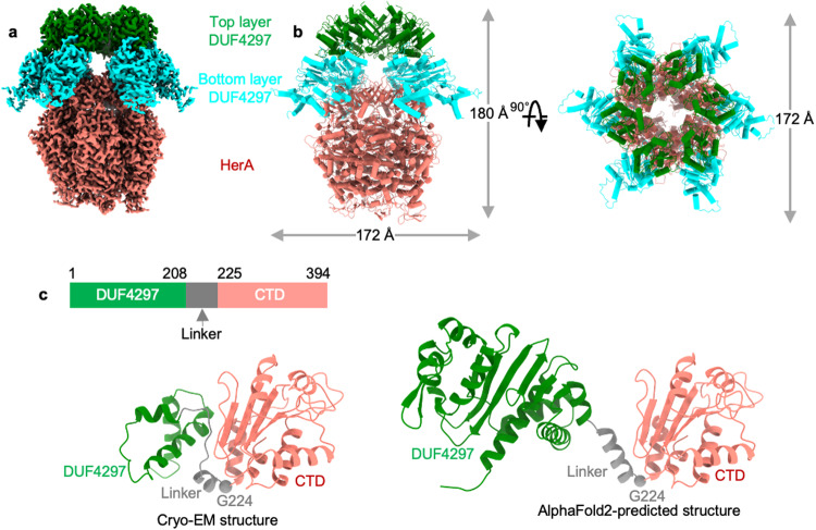
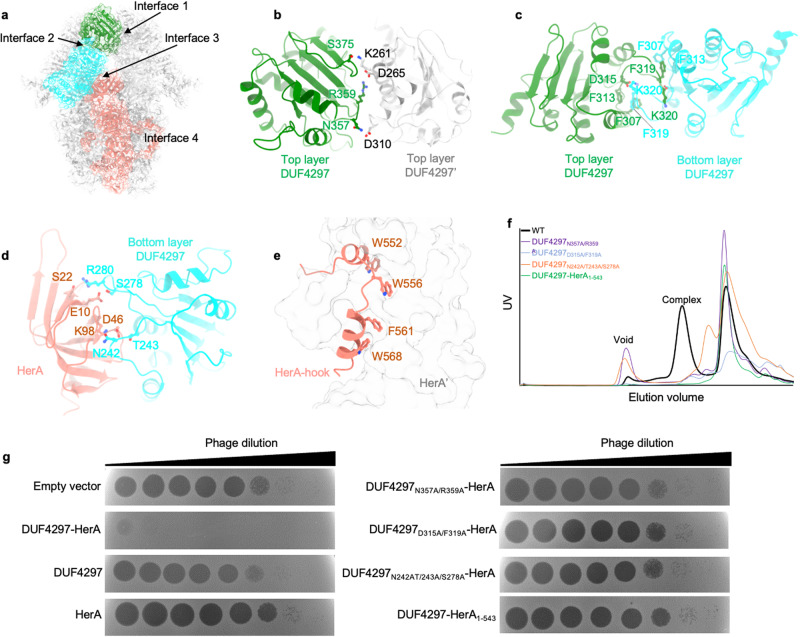
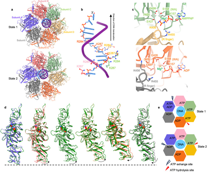
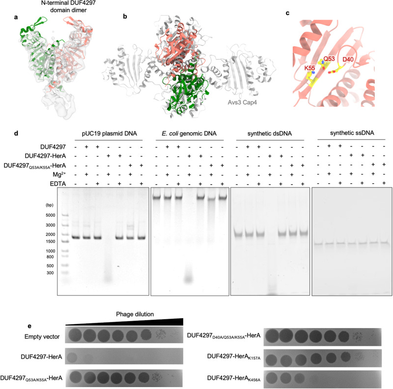
Coupling distinct enzymatic effectors emerges as an efficient strategy for defense against phage infection in bacterial immune responses, such as the widely studied nuclease and cyclase activities in the type III CRISPR-Cas system. However, concerted enzymatic activities in other bacterial defense systems are poorly understood. Here, we biochemically and structurally characterize a two-component defense system DUF4297-HerA, demonstrating that DUF4297-HerA confers resistance against phage infection by cooperatively cleaving dsDNA and hydrolyzing ATP. DUF4297 alone forms a dimer, and HerA alone exists as a nonplanar split spiral hexamer, both of which exhibit extremely low enzymatic activity. Interestingly, DUF4297 and HerA assemble into an approximately 1 MDa supramolecular complex, where two layers of DUF4297 (6 DUF4297 molecules per layer) linked via inter-layer dimerization of neighboring DUF4297 molecules are stacked on top of the HerA hexamer. Importantly, the complex assembly promotes dimerization of DUF4297 molecules in the upper layer and enables a transition of HerA from a nonplanar hexamer to a planar hexamer, thus activating their respective enzymatic activities to abrogate phage infection. Together, our findings not only characterize a novel dual-enzyme anti-phage defense system, but also reveal a unique activation mechanism by cooperative complex assembly in bacterial immunity.


Conflict of interest statement

The authors declare no competing interests.

Figures

Fig. 1
Fig. 1. Cryo-EM structure of HerA hexamer.
a Cryo-EM density map of HerA. b Overall structure of HerA. c Domain organization of HerA. Each domain is labeled and in a different color. The structure of SsoHerA (PDB: 4D2I) is shown for comparison. d ATPase activities of HerA and DUF4297–HerA complex.
Fig. 2
Fig. 2. Cryo-EM structure of the DUF4297–HerA complex.
a Cryo-EM density map of DUF4297–HerA complex. b Overall structure of the DUF4297–HerA complex. HerA, the bottom layer DUF4297, and the top layer DUF4297 are colored salmon, cyan, and green, respectively. c Domain organization of DUF4297. Each domain is labeled and color-coded. The AlphaFold2-predicted structure of DUF4297 is shown for comparison.
Fig. 3
Fig. 3. Assembly of DUF4297–HerA complex.
a Four key assembly interfaces of the DUF4297–HerA complex are indicated. b Detailed interactions between the CTDs of two adjacent upper layer DUF4297 molecules. c Detailed interactions between the CTDs of the upper layer DUF4297 and the corresponding bottom layer DUF4297. d Detailed interactions between the CTD of the bottom layer DUF4297 and the HAS domain of HerA. e Detailed interactions between the C-terminal hook and the adjacent HerA. f Size-exclusion chromographs of DUF4297–HerA wild type (WT) and mutants harboring mutations at the assembly interface. g Plaques of phage λ on cells expressing empty vector, DUF4297–HerA WT, and the indicated mutants. 10-fold serial dilutions of the phage lysate were dropped on the plates.
Fig. 4
Fig. 4. Cryo-EM structures of DUF4297–HerA complexed with ATP analogs and DNA.
a Bottom view of DUF4297–HerA complexed with ATP analogs and DNA in state 1 (upper panel) and state 2 (lower panel). DUF4297 is removed for clarity. b Interactions between DNA and the two basic residues of four DNA-engaged subunits in state 1. c Detailed interactions between HerA and the phosphate groups of AMPPNP (upper panel) and ADP (lower panel). d Conformational changes of each HerA subunit between state 1 and state 2. The HerA subunit of state 1 is color-coded as in a, while the HerA subunit of state 2 is colored green. e Nucleotide-binding status in state 1 (upper panel) and state 2 (lower panel).
Fig. 5
Fig. 5. Dimerization of the N-terminal DUF4297 domain.
a Orthogonal views of the predicted N-terminal domain fitted into the 4.2 Å local-refined cryo-EM map. b Superimposition of the N-terminal DUF4297 domain dimer and the Avs3 cap4 domain tetramer. c Active site of the N-terminal DUF4297 domain. d Agarose gel analysis of the nuclease activity of the DUF4297–HerA complex in vitro with a pUC19 plasmid DNA, E. coli genomic DNA, synthetic dsDNA, or synthetic ssDNA. e Plaques of phage λ on cells expressing empty vector, DUF4297–HerA WT, and the indicated mutants. 10-fold serial dilutions of the phage lysate were dropped on the plates.
Fig. 6
Fig. 6. Mechanism of anti-phage defense by DUF4297–HerA.
a Bacterial growth curves in the presence of DUF4297–HerA upon infection by phage λ with the indicated MOI values. b Bacterial growth curves in the presence of DUF4297–HerA mutants (DUF4297mut: DUF4297D40A/Q53A/K55A–HerA; HerAmut: DUF4297–HerAK157A) upon infection by phage λ with the indicated MOI values. c Representative images of cells expressing DUF4297–HerA or mutants either uninfected or 60 min post infection with phage λ at an MOI of 1. Cell membranes were stained with FM4-64 (red), and DNA was stained with DAPI (blue). White arrows indicate the ‘phantom’ cells devoid of both phage and host DNA. d Quantification of the percentage of ‘phantom’ cells in c. e Assembly and possible activation mechanisms of the DUF4297–HerA complex.

References

    1. Hendrix, R. W., Smith, M. C. M., Burns, R. N., Ford, M. E. & Hatfull, G. F. Evolutionary relationships among diverse bacteriophages and prophages: All the world’s a phage. Proc. Natl. Acad. Sci. USA96, 2192–2197 (1999). - PMC - PubMed
    1. Simmonds, P. et al. Virus taxonomy in the age of metagenomics. Nat. Rev. Microbiol.15, 161–168 (2017). - PubMed
    1. Tesson, F. et al. Systematic and quantitative view of the antiviral arsenal of prokaryotes. Nat. Commun.13, 2561 (2022). - PMC - PubMed
    1. Payne, L. J. et al. Identification and classification of antiviral defence systems in bacteria and archaea with PADLOC reveals new system types. Nucleic Acids Res.49, 10868–10878 (2021). - PMC - PubMed
    1. Doron, S. et al. Systematic discovery of antiphage defense systems in the microbial pangenome. Science359, eaar4120 (2018). - PMC - PubMed

MeSH terms

LinkOut - more resources