

Retinal blood flow association with age and weight in infants at risk for retinopathy of prematurity
- PMID: 38834830
- PMCID: PMC11150459
- DOI: 10.1038/s41598-024-63534-6
Retinal blood flow association with age and weight in infants at risk for retinopathy of prematurity
Abstract
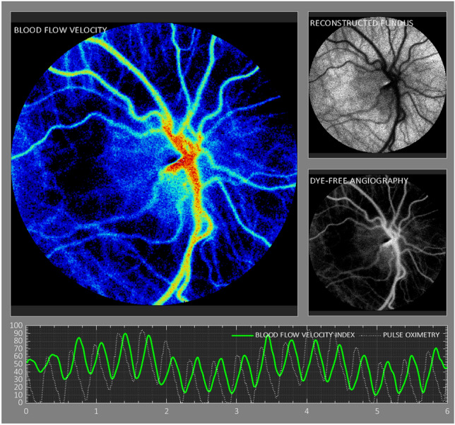
This prospective study evaluated the relationship between laser speckle contrast imaging (LSCI) ocular blood flow velocity (BFV) and five birth parameters: gestational age (GA), postmenstrual age (PMA) and chronological age (CA) at the time of measurement, birth weight (BW), and current weight (CW) in preterm neonates at risk for retinopathy of prematurity (ROP). 38 Neonates with BW < 2 kg, GA < 32 weeks, and PMA between 27 and 47 weeks underwent 91 LSCI sessions. Correlation tests and regression analysis were performed to quantify relationships between birth parameters and ocular BFV. Mean ocular BFV index in this cohort was 8.8 +/- 4.0 IU. BFV positively correlated with PMA (r = 0.3, p = 0.01), CA (r = 0.3, p = 0.005), and CW (r = 0.3, p = 0.02). BFV did not correlate with GA nor BW (r = - 0.2 and r = - 0.05, p > 0.05). Regression analysis with mixed models demonstrated that BFV increased by 1.2 for every kilogram of CW, by 0.34 for every week of CA, and by 0.36 for every week of PMA (p = 0.03, 0.004, 0.007, respectively). Our findings indicate that increased age and weight are associated with increased ocular BFV measured using LSCI in premature infants. Future studies investigating the associations between ocular BFV and ROP clinical severity must control for age and/or weight of the infant.
© 2024. The Author(s).
Conflict of interest statement
Dr. Alexander’s work has been funded by Maryland Industrial Partnerships (MIPS) Program (Grant 7103, funded in part by Vasoptic Medical). The remaining authors declare no competing interests.
Figures

Update of
-
Retinal blood flow association with age and weight in infants at risk for retinopathy of prematurity.Res Sq [Preprint]. 2024 Feb 22:rs.3.rs-3909449. doi: 10.21203/rs.3.rs-3909449/v1. Res Sq. 2024. Update in: Sci Rep. 2024 Jun 4;14(1):12790. doi: 10.1038/s41598-024-63534-6. PMID: 38464120 Free PMC article. Updated. Preprint.
References
MeSH terms
Grants and funding
LinkOut - more resources
Full Text Sources
Medical
