Review
. 2024 Jun;14(6):240041.
doi: 10.1098/rsob.240041. Epub 2024 Jun 5.

A circle of life: platelet and megakaryocyte cytoskeleton dynamics in health and disease

Affiliations
Review

A circle of life: platelet and megakaryocyte cytoskeleton dynamics in health and disease

Haonan Liu et al. Open Biol. 2024 Jun.

Abstract

Platelets are blood cells derived from megakaryocytes that play a central role in regulating haemostasis and vascular integrity. The microtubule cytoskeleton of megakaryocytes undergoes a critical dynamic reorganization during cycles of endomitosis and platelet biogenesis. Quiescent platelets have a discoid shape maintained by a marginal band composed of microtubule bundles, which undergoes remarkable remodelling during platelet activation, driving shape change and platelet function. Disrupting or enhancing this process can cause platelet dysfunction such as bleeding disorders or thrombosis. However, little is known about the molecular mechanisms underlying the reorganization of the cytoskeleton in the platelet lineage. Recent studies indicate that the emergence of a unique platelet tubulin code and specific pathogenic tubulin mutations cause platelet defects and bleeding disorders. Frequently, these mutations exhibit dominant negative effects, offering valuable insights into both platelet disease mechanisms and the functioning of tubulins. This review will highlight our current understanding of the role of the microtubule cytoskeleton in the life and death of platelets, along with its relevance to platelet disorders.

Keywords: megakaryocyte; microtubule; motors; pathogenesis; platelet; tubulin.


Conflict of interest statement

We declare we have no competing interests.

Figures

Structural location of the most frequent tubulin mutations associated with platelet disorders mapped onto a tubulin heterodimer
Figure 1.
Structural location of the most frequent tubulin mutations associated with platelet disorders mapped onto a tubulin heterodimer. Amino acid residues in which frequent mutations have been identified to cause platelet disorders are highlighted (a) β1-tubulin (b) α4-tubulin and (c) α8-tubulin in different colours (α-tubulin in white and β-tubulin in brown). Orange and green sticks show GTP/GDP nucleotide on α- and β-tubulins, respectively. The three-dimensional structure of tubulin heterodimers was visualized by PyMOL using PDB ID 1JFF.
Schematic of microtubule function during in vitro and in vivo platelet formation from megakaryocytes
Figure 2.
Schematic of microtubule function during in vitro and in vivo platelet formation from megakaryocytes. (a) Megakaryocytes differentiate from haematopoietic stem cells within the bone marrow, which is regulated by thrombopoietin (TPO). (b) Megakaryocytes undergo endomitosis and maturation, resulting in polyploidy, enlarged cell size and platelet-specific protein synthesis. Microtubules are assembled into multiple spindles that support asymmetric chromosome segregation. (c) The sliding of spindle microtubules drives centrosome clustering within a typical 16N DNA mature megakaryocyte. A monospindle emanates from the microtubule-organizing centre to the cell cortex. (d) Before proplatelet formation, megakaryocyte centrosomes cluster into a microtubule-organizing centre, and microtubules relocate to the cell cortex. (e) Microtubule bundles are cross-linked by microtubule-associated proteins formed beneath the pseudopodia. Sliding of overlapping microtubules by dynein results in proplatelet elongation. Kinesins transport mitochondria, granules and other organelles into proplatelet far ends and are trapped with the microtubule coil. (f) Models for platelet release from megakaryocytes. In the in vitro model, megakaryocytes migrate to vascular sinusoids; elongated proplatelets are released into the bloodstream and fragmented into large discoid preplatelets. Preplatelets interconvert into barbell-shaped proplatelets under shear stress—dynamic microtubule reorganization by twisting, merging and folding causes barbell-shaped proplatelets to split into two platelets. In the in vivo model, megakaryocytes release large protrusions into sinusoid blood, then fragment into proplatelets or preplatelets in the microcirculation. Figure created using BioRender.
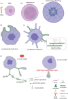
Schematic of the marginal band microtubule reorganization during platelet activation
Figure 3.
Schematic of marginal band microtubule reorganization during platelet activation. (a) Discoid platelets contain a circumferential marginal band of multiple dynamic short microtubules and a stable long microtubule, kept at equilibrium owing to kinesin antagonizing dynein motor activity microtubule-associated proteins cross-linking microtubules. (b) Activation of platelets causes inhibition of kinesin activity, in which dynein starts to slide microtubules, resulting in elongation and coiling of the marginal band, leading to compression of the actin cortex and the disc-to-sphere shape change. Polymerization of microtubule and actin filaments results in the formation of filopodia-like cell protrusions that facilitate platelet adhesion and aggregation. Figure created using BioRender.

Similar articles

References

    1. Cimmino G, Golino P. 2013. Platelet biology and receptor pathways. J. Cardiovasc. Transl. Res. 6, 299–309. (10.1007/s12265-012-9445-9) - DOI - PubMed
    1. Josefsson EC, Vainchenker W, James C. 2020. Regulation of platelet production and lifespan: role of Bcl-xL and potential implications for human platelet diseases. Int. J. Mol. Sci. 21, 7591. (10.3390/ijms21207591) - DOI - PMC - PubMed
    1. Lebois M, Josefsson EC. 2016. Regulation of platelet lifespan by apoptosis. Platelets 27, 497–504. (10.3109/09537104.2016.1161739) - DOI - PubMed
    1. Lefrançais E, Looney MR. 2019. Platelet biogenesis in the lung circulation. Physiology (Bethesda). 34, 392–401. (10.1152/physiol.00017.2019) - DOI - PMC - PubMed
    1. White JG, Rao GH. 1998. Microtubule coils versus the surface membrane cytoskeleton in maintenance and restoration of platelet discoid shape. Am. J. Pathol. 152, 597–609. - PMC - PubMed

LinkOut - more resources