. 2024 May;62(2):243-250.
doi: 10.3347/PHD.23125. Epub 2024 May 27.

Organ-specific Toxocara canis larvae migration and host immune response in experimentally infected mice

Affiliations

Organ-specific Toxocara canis larvae migration and host immune response in experimentally infected mice

Min Seok Kim et al. Parasites Hosts Dis. 2024 May.

Abstract

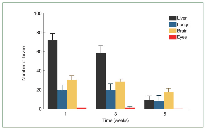
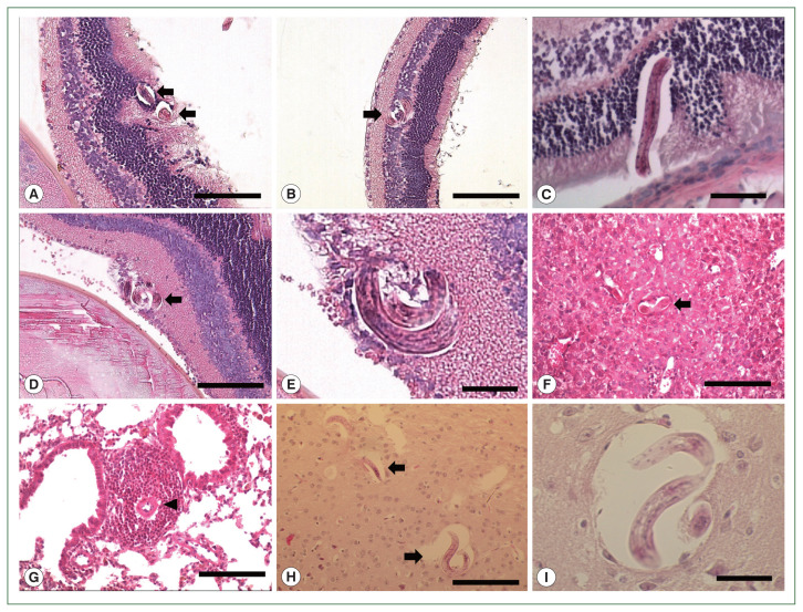
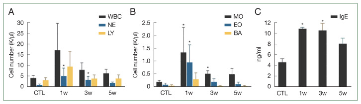
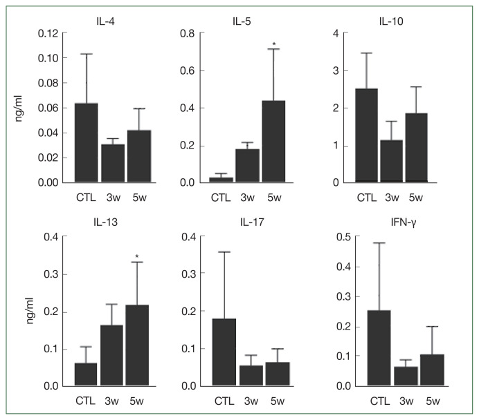
We investigated organ specific Toxocara canis larval migration in mice infected with T. canis larvae. We observed the worm burden and systemic immune responses. Three groups of BALB/c mice (n=5 each) were orally administered 1,000 T. canis 2nd stage larvae to induce larva migrans. Mice were sacrificed at 1, 3, and 5 weeks post-infection. Liver, lung, brain, and eye tissues were collected. Tissue from 2 mice per group was digested for larval count, while the remaining 3 mice underwent histological analysis. Blood hematology and serology were evaluated and compared to that in a control uninfected group (n=5) to assess the immune response. Cytokine levels in bronchoalveolar lavage (BAL) fluid were also analyzed. We found that, 1 week post-infection, the mean parasite load in the liver (72±7.1), brain (31±4.2), lungs (20±5.7), and eyes (2±0) peaked and stayed constant until the 3 weeks. By 5-week post-infection, the worm burden in the liver and lungs significantly decreased to 10±4.2 and 9±5.7, respectively, while they remained relatively stable in the brain and eyes (18±4.2 and 1±0, respectively). Interestingly, ocular larvae resided in all retinal layers, without notable inflammation in outer retina. Mice infected with T. canis exhibited elevated levels of neutrophils, monocytes, eosinophils, and immunoglobulin E. At 5 weeks post-infection, interleukin (IL)-5 and IL-13 levels were elevated in BAL fluid. Whereas IL-4, IL-10, IL-17, and interferon-γ levels in BAL fluid were similar to that in controls. Our findings demonstrate that a small portion of T. canis larvae migrate to the eyes and brain within the first week of infection. Minimal tissue inflammation was observed, probably due to increase of anti-inflammatory cytokines. This study contributes to our understanding of the histological and immunological responses to T. canis infection in mice, which may have implications to further understand human toxocariasis.

Keywords: Toxocara canis; host-parasite interaction; larva migrans; toxocariasis.


Conflict of interest statement

Conflict of interest: The authors have no potential conflict of interest to disclosure.

Figures

Fig. 1
Fig. 1
Migratory behavior of Toxocara canis larvae in BALB/c mice. Mean number of total Toxocara canis larvae recovered from 4 organs of BALB/c infected with a 1,000-larvae dose after 1-, 3-, and 5-week post-infection.
Fig. 2
Fig. 2
Histopathological findings of the Toxocara canis larvae infected tissue. Toxocara canis larvae (arrows) in the retina (A–E), liver (F), lung (G), and brain (H and I) in the first week post-infection. Note that Toxocara canis larvae are present in all layers of retina. Compared to the larvae in the outer or middle retina (A–C), the larvae in the inner retina (D and E) recruit multiple eosinophils in the vitreous and the inner surface of the retina. Eosinophil infiltration (arrowhead) was markedly noted in the lung (G) (H & E, 200× magnification; Scale bar: 100 μm). Magnified images of larvae in the retina (C and E) and brain (I) (400× magnification; Scale bar: 20 μm).
Fig. 3
Fig. 3
Changes in blood cell count and antibodies in blood in response to Toxocara canis larval infection. Differential blood cell counts in control and infected mice (A, B); Serum IgE titers were higher in infected mice than control mice at 1, 3, and 5 weeks (C). Significant differences compared to control group are indicated by an asterisk (P<0.017, Kruskal–Wallis test). CTL, control mice; INF, infected mice; NE, neutrophil; LY, lymphocyte; MO, monocyte; EO, eosinophil; BA, basophil.
Fig. 4
Fig. 4
Cytokines in bronchoalveolar lavage fluid (BAL). Significant differences compared to control group are indicated by an asterisk (P<0.025, Kruskal–Wallis test). CTL, control; INF, infected mice.

Similar articles

References

    1. Ahn SJ, Ryoo NK, Woo SJ. Ocular toxocariasis: clinical features, diagnosis, treatment, and prevention. Asia Pac Allergy. 2014;4(3):134–141. doi: 10.5415/apallergy.2014.4.3.134. - DOI - PMC - PubMed
    1. Kolbekova P, Kolarova L, Vetvicka D, Syrucek M. Imaging of Toxocara canis larvae labelled by CFSE in BALB/c mice. Parasitol Res. 2011;108(4):1007–1014. doi: 10.1007/s00436-010-2145-y. - DOI - PubMed
    1. Beaver PC, Snyder CH, Carrera GM, Dent JH, Lafferty JW. Chronic eosinophilia due to visceral larva migrans; report of three cases. Pediatrics. 1952;9(1):7–19. doi: 10.1542/peds.9.1.7. - DOI - PubMed
    1. Yang HK, Woo SJ, Hwang JM. Toxocara optic neuropathy after ingestion of raw meat products. Optom Vis Sci. 2014;91(11):e267–273. doi: 10.1097/OPX.0000000000000404. - DOI - PubMed
    1. Strube C, Heuer L, Janecek E. Toxocara spp. infections in paratenic hosts. Vet Parasitol. 2013;193(4):375–389. doi: 10.1016/j.vetpar.2012.12.033. - DOI - PubMed

MeSH terms

LinkOut - more resources