. 2024 May 20:15:1342444.
doi: 10.3389/fmicb.2024.1342444. eCollection 2024.

Genome-wide CRISPR/Cas9 screen reveals JunB downmodulation of HIV co-receptor CXCR4

Affiliations

Genome-wide CRISPR/Cas9 screen reveals JunB downmodulation of HIV co-receptor CXCR4

William J Schulze et al. Front Microbiol. .

Abstract

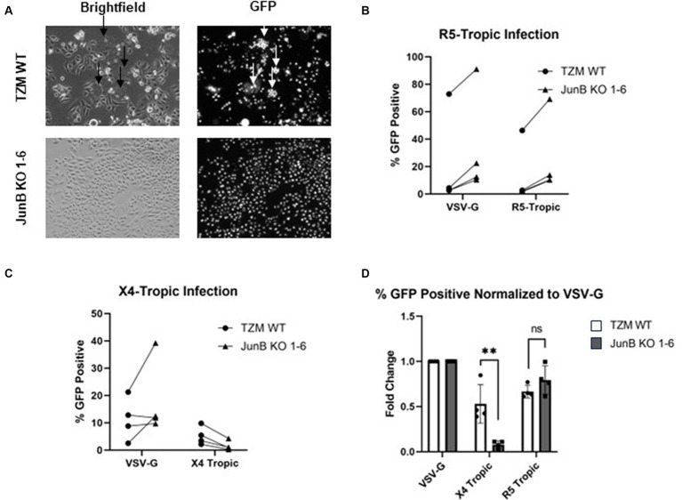
HIV-1 relies extensively on host cell machinery for replication. Identification and characterization of these host-virus interactions is vital to our understanding of viral replication and the consequences of infection in cells. Several prior screens have identified host factors important for HIV replication but with limited replication of findings, likely due to differences in experimental design and conditions. Thus, unidentified factors likely exist. To identify novel host factors required for HIV-1 infection, we performed a genome-wide CRISPR/Cas9 screen using HIV-induced cell death as a partitioning method. We created a gene knockout library in TZM-GFP reporter cells using GeCKOv2, which targets 19,050 genes, and infected the library with a lethal dose of HIV-1NL4-3. We hypothesized that cells with a knockout of a gene critical for HIV infection would survive while cells with a knockout of a non-consequential gene would undergo HIV-induced death and be lost from the population. Surviving cells were analyzed by high throughput sequencing of the integrated CRISPR/Cas9 cassette to identify the gene knockout. Of the gene targets, an overwhelming majority of the surviving cells harbored the guide sequence for the AP-1 transcription factor family protein, JunB. Upon the generation of a clonal JunB knockout cell line, we found that HIV-1NL4-3 infection was blocked in the absence of JunB. The phenotype resulted from downregulation of CXCR4, as infection levels were recovered by reintroduction of CXCR4 in JunB KO cells. Thus, JunB downmodulates CXCR4 expression in TZM-GFP cells, reducing CXCR4-tropic HIV infection.

Keywords: CRISPR/Cas9; CXCR4; HIV; JunB; host-factors.


Conflict of interest statement

The authors declare that the research was conducted in the absence of any commercial or financial relationships that could be construed as a potential conflict of interest.

Figures

Figure 1
Figure 1
Genome-wide CRISPR/Cas9 screen. (A) Schematic of LentiCRISPRv2 HIV host factor screen. Cells were transduced with the LentiCRISPRv2 library (Addgene) at an MOI of 0.1. Transduced cells that survived puromycin selection and continued to proliferate were subjected to a lethal dose of HIV-1NL4-3. Surviving cells were subjected to DNA isolation, amplification of the gRNA cassette, and regeneration of the LentiCRISPRv2 library (R1). After two rounds of selection with a lethal dose of HIV-1NL4-3 (MOI ~3), the surviving cell population (R2) was subjected to DNA isolation, PCR amplification of the gRNA cassette with Illumina adapters, and high throughput sequencing to identify edited genes. (B) Percentage of the total reads of individual gRNAs from HTS sequencing conducted on cells that survived two rounds of selection with a lethal dose of HIV-1NL4-3. (C) Comparison of normalized reads in both the R2 and starting populations by HTS. Each dot represents a single gRNA. Dots above the dotted line (x = y) were enriched in the selected population, while those below were depleted. (D) The difference in normalized reads from the starting library to the R2 selected library for individual genes.
Figure 2
Figure 2
Polyclonal JunB KO cells remained susceptible to HIV-1NL4-3 infection but resistant to HIV-induced cell death. (A) Representative brightfield and GFP fluorescence microscopy images of TZM WT cells and JunB KO polyclonal cells infected with a lethal dose of HIV-1NL4-3 (48-h timepoint). GFP-positive cells are presumed to be infected with HIV-1NL4-3. Arrows indicate the presence of syncytia. (B) TZM WT cells and JunB KO polyclonal cells were plated in 96-well plates and infected with a lethal dose of HIV-1NL4-3 (MOI ~ 3). After 72 h, viability was determined using the CellTiter96 kit. (C) Flow cytometry analysis of TZM WT and TZM-GFP JunB KO polyclonal cells infected with dilutions of HIV-1NL4-3 in 12-well plates. Error bars represent standard deviation of biological replicates, n = 3 (Student’s t-test, ****p < 0.0001, *p < 0.05).
Figure 3
Figure 3
TZM-GFP JunB KO 1–6 cells are resistant to infection of X4-tropic virus. (A) Representative brightfield and GFP fluorescence microscopy images of TZM WT and TZM-GFP JunB 1–6 KO cells infected with Env-encoding HIV-1 pseudotyped with VSV-G. Arrows indicate syncytia formation. (B) Flow cytometry analysis on TZM WT or TZM-GFP JunB KO 1–6 cells infected with single cycle HIV-1 pseudotyped with VSV-G or R5-tropic Bal envelope. Lines indicate paired replicates where the same virus prep was used to infect the two cell lines. (C) Flow cytometry analysis on TZM WT or TZM-GFP JunB KO 1–6 cells infected with single cycle HIV-1 pseudotyped with VSV-G or X4-tropic NL4-3 envelope. Lines indicate paired replicates where the same virus prep was used to infect the two cell lines. (D) Normalized flow cytometry analysis on TZM WT or TZM-GFP JunB KO 1–6 cells infected with single cycle HIV-1 pseudotyped with VSV-G, X4-tropic NL4-3 envelope, and R5-tropic Bal envelope. Represented as percent infected cells normalized to percent infected in VSV-G infection. Error bars represent standard deviation of biological replicates (Student’s t-test, **p < 0.01).
Figure 4
Figure 4
TZM-GFP JunB KO cells have decreased CXCR4 mRNA and protein expression. Flow cytometry analysis of cell surface expression of HIV receptor (A) CD4, and (B) co-receptor CXCR4 using PE-labeled antibodies. qPCR analyses on mRNA isolated from TZM WT and TZM-GFP JunB KO 1–6 cells for (C) CXCR4 mRNA and (D) CCR5 mRNA normalized to TZM WT GAPDH mRNA. Each point in (C,D) are biological replicates, error bars represent standard deviation. Statistical analysis conducted on non-normalized dCT value as shown in Supplementary Figure S4 (Wilcoxon matched-pairs signed rank test, **p < 0.01). Experiments were repeated three times.
Figure 5
Figure 5
Transfection of exogenous CXCR4 restored the infectivity of X4-tropic HIV. (A) Transfection of plasmid pcDNA3.1-CXCR4 restored the surface expression of CXCR4 by flow cytometry using PE-conjugated antibody. A representative experiment of three total experiments is shown. (B) Non-normalized flow cytometry analysis of TZM WT or TZM-GFP JunB KO 1–6 cells transfected with either control plasmid pcDNA3.1-empty or plasmid pcDNA3.1-CXCR4 and infected with single cycle HIV-1 pseudotyped with VSV-G. Lines indicate paired replicates where the same virus prep was used to infect the indicated cells. (C) Non-normalized flow cytometry analysis of TZM WT or TZM-GFP JunB KO 1–6 cells transfected with either control plasmid pcDNA3.1-empty or plasmid pcDNA3.1-CXCR4 and infected with single cycle HIV-1 pseudotyped with X4-tropic NL4-3 envelope. (D) Flow cytometry analysis of cells transfected with control plasmid pcDNA3.1-empty or plasmid pcDNA3.1-CXCR4 and infected with single cycle HIV-1 with the indicated glycoprotein. Represented as percent infected normalized to percent infected in VSV-G infections. Error bars represent standard deviation of biological replicates, n = 7. Kruskal–Wallis test, ***p < 0.001.
Figure 6
Figure 6
Promoter schematics and representative images of 2% agarose gel electrophoresis of PCR products corresponding to the promoter region of (A) CXCR4, (B) CD4, and (C) CCR5 using DNA recovered from chromatin immunoprecipitation assays in TZM WT, TZM-GFP JunB KO 1–6 (JBKO), and CEM T4 (CEM) cells.

Similar articles

References

    1. Alam K. K., Chang J. L., Burke D. H. (2015). FASTAptamer: a bioinformatic toolkit for high-throughput sequence analysis of combinatorial selections. Mol. Ther. Nucleic Acids 4:e230. doi: 10.1038/mtna.2015.4, PMID: - DOI - PMC - PubMed
    1. Arthos J., Deen K. C., Chaikin M. A., Fornwald J. A., Sathe G., Sattentau Q. J., et al. . (1989). Identification of the residues in human CD4 critical for the binding of HIV. Cell 57, 469–481. doi: 10.1016/0092-8674(89)90922-7, PMID: - DOI - PubMed
    1. Balkhi M. Y., Wittmann G., Xiong F., Junghans R. P. (2018). YY1 upregulates checkpoint receptors and downregulates type I cytokines in exhausted, chronically stimulated human T cells. iScience. 2, 105–122. doi: 10.1016/j.isci.2018.03.009, PMID: - DOI - PMC - PubMed
    1. Bazan H. A., Alkhatib G., Broder C. C., Berger E. A. (1998). Patterns of CCR5, CXCR4, and CCR3 usage by envelope glycoproteins from human immunodeficiency virus type 1 primary isolates. J. Virol. 72, 4485–4491. doi: 10.1128/JVI.72.5.4485-4491.1998, PMID: - DOI - PMC - PubMed
    1. Berger E. A., Doms R. W., Fenyö E. M., Korber B. T. M., Littman D. R., Moore J. P., et al. . (1998). A new classification for HIV-1. Nature 391:240. doi: 10.1038/34571 - DOI - PubMed