Review
. 2024 May 21:12:1391259.
doi: 10.3389/fcell.2024.1391259. eCollection 2024.

The influence of viscosity of hydrogels on the spreading and migration of cells in 3D bioprinted skin cancer models

Affiliations
Review

The influence of viscosity of hydrogels on the spreading and migration of cells in 3D bioprinted skin cancer models

Lissinda H Du Plessis et al. Front Cell Dev Biol. .

Abstract

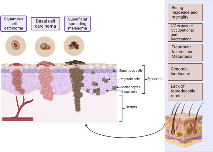
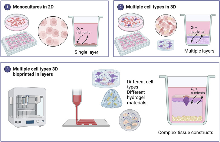
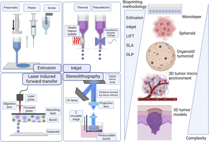
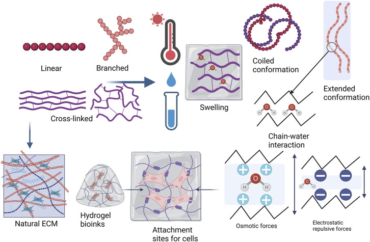
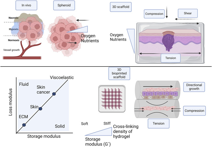
Various in vitro three-dimensional (3D) tissue culture models of human and diseased skin exist. Nevertheless, there is still room for the development and improvement of 3D bioprinted skin cancer models. The need for reproducible bioprinting methods, cell samples, biomaterial inks, and bioinks is becoming increasingly important. The influence of the viscosity of hydrogels on the spreading and migration of most types of cancer cells is well studied. There are however limited studies on the influence of viscosity on the spreading and migration of cells in 3D bioprinted skin cancer models. In this review, we will outline the importance of studying the various types of skin cancers by using 3D cell culture models. We will provide an overview of the advantages and disadvantages of the various 3D bioprinting technologies. We will emphasize how the viscosity of hydrogels relates to the spreading and migration of cancer cells. Lastly, we will give an overview of the specific studies on cell migration and spreading in 3D bioprinted skin cancer models.

Keywords: 3D bioprinting; cell interaction; hydrogels; melanoma; skin cancer.


Conflict of interest statement

The authors declare that the research was conducted in the absence of any commercial or financial relationships that could be construed as a potential conflict of interest.

Figures

FIGURE 1
FIGURE 1
The most common types of skin cancer within the different layers of the skin. Squamous cell carcinoma arises from the squamous cells and basal cell carcinoma from the basal cell layer, these cells form the epidermal layer of the skin. Melanoma arises from melanocytes found in the basal cell layer. Melanoma has two major phases of progression. In the radial growth phase that takes place in the superficial layers of the skin, lesions are recognized as a pigmented area or plaque. In the vertical growth phase, the tumor may elevate the epidermis to give the appearance of a nodule, or it may penetrate the dermis. There may be individual abnormal melanocytes or small clusters of these cells present known as pagetoid cells or pagetoid spread of melanoma. Some of the most important challenges in skin cancer research are listed. Created with BioRender.com.
FIGURE 2
FIGURE 2
Summary of the different models used in skin cancer research. Monocultures of skin cancer cells include one cell type grown in a single layer. Various methods of 3D cell culture exist including scaffold-free techniques such as spheroids, organoids, and skin-on-chip., Scaffold-based 3D models include a hydrogel matrix. These models can be monocultures but for skin cancer, most 3D models include multiple cell types grown in layers to represent the in vivo skin more accurately. 3D bioprinting offers a unique advantage in being able to combine different cell types in different hydrogels (biomaterial inks) to create bioinks (hydrogel biomaterials combined with cells) for a fabricated skin model. These 3D skin cancer models can be biofabricated in a layer-by-layer approach to create complex tissue constructs more accurately. Created with BioRender.com.
FIGURE 3
FIGURE 3
Evolution of skin cancer models and technologies from scaffold-free 3D structures to more complex 3D structures. Laser-induced forward transfer (LIFT), stereolithography (SLA), and digital light processing (DLP). Created with BioRender.com.
FIGURE 4
FIGURE 4
Hydrogel material polymer properties and topology, describing whether a polymer structure is linear, branched, cross-linked, or a network. Hydrogels tend to obtain freedom of movement of the polymer chains as they are placed in water or biological fluids and when temperature is increased. Swelling in a hydrogel is generally determined by polymer-solvent interaction in nonionic hydrogels and by osmotic or electrostatic repulsive forces if the hydrogel is ionic. Higher cross-linking density leads to decreased mesh size or porosity with increased stiffness. The polymer chains in the hydrogel bioink resemble the natural ECM and the swelled hydrogel provides attachment sites for cells (Adapted from Berger et al., 2004; Mehta et al., 2023; Sinko, 2011; Xu J et al., 2022a). Created with BioRender.com.
FIGURE 5
FIGURE 5
Comparison of different types of 3D skin cancer models with in vivo conditions, focusing on scaffold-free, scaffold-based, and 3D bioprinted scaffolds in multiple layers. To facilitate optimal growth and proliferation of cells in 3D models, oxygen and nutrient supply is important. Cell-cell interaction is influenced by mechanical forces and tissue stiffness, including compression, tension, and shear stresses. Tissue stiffness of the native ECM, skin, and skin tumor differ and should be incorporated in the skin cancer models. 3D bioprinting in layers confers directional growth. The hydrogel in the multilayer 3D bioprinted scaffold provides osmotic and hydrostatic pressure. Created with BioRender.com.

Similar articles

Cited by

References

    1. Agache P. G., Monneur C., Leveque J.-L., De Rigai J. (1980). Mechanical properties and Young's modulus of human skin in vivo . Arch. Dermatol. Res. 269, 221–232. 10.1007/BF00406415 - DOI - PubMed
    1. Agrawal R., Woodfolk J. A. (2014). Skin barrier defects in atopic dermatitis. Curr. Allergy Asthma Rep. 14, 433. 10.1007/s11882-014-0433-9 - DOI - PMC - PubMed
    1. Ahmed E. M. (2015). Hydrogel: preparation, characterization, and applications: a review. J. Adv. Res. 6 (2), 105–121. 10.1016/j.jare.2013.07.006 - DOI - PMC - PubMed
    1. Ahn G., Min K. H., Kim C., Lee J. S., Kang D., Won J. Y., et al. (2017). Precise stacking of decellularized extracellular matrix based 3D cell-laden constructs by a 3D cell printing system equipped with, heating modules. Sci. Rep-Uk 7 (1), 8624. 10.1038/s41598-017-09201-5 - DOI - PMC - PubMed
    1. Ahn M., Cho W. W., Park W., Lee J. S., Choi M. J., Gao Q., et al. (2023). 3D biofabrication of diseased human skin models in vitro . Biomater. Res. 27 (1), 80. 10.1186/s40824-023-00415-5 - DOI - PMC - PubMed

LinkOut - more resources