. 2024 May 21:12:1411162.
doi: 10.3389/fcell.2024.1411162. eCollection 2024.

Gene-deficient mouse model established by CRISPR/Cas9 system reveals 15 reproductive organ-enriched genes dispensable for male fertility

Affiliations

Gene-deficient mouse model established by CRISPR/Cas9 system reveals 15 reproductive organ-enriched genes dispensable for male fertility

Tuyen Thi Thanh Nguyen et al. Front Cell Dev Biol. .

Abstract

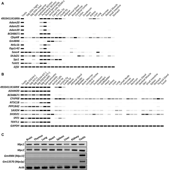
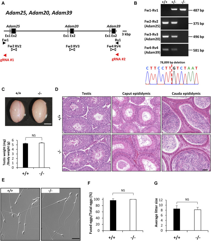
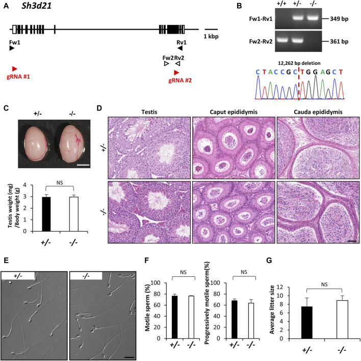
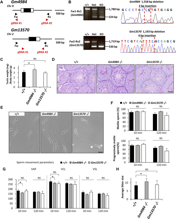
Since the advent of gene-targeting technology in embryonic stem cells, mice have become a primary model organism for investigating human gene function due to the striking genomic similarities between the two species. With the introduction of the CRISPR/Cas9 system for genome editing in mice, the pace of loss-of-function analysis has accelerated significantly. This has led to the identification of numerous genes that play crucial roles in male reproductive processes, including meiosis, chromatin condensation, flagellum formation in the testis, sperm maturation in the epididymis, and fertilization in the oviduct. Despite the advancements, the functions of many genes, particularly those enriched in male reproductive tissues, remain largely unknown. In our study, we focused on 15 genes and generated 13 gene-deficient mice [4933411K16Rik, Adam triple (Adam20, Adam25, and Adam39), BC048671, Cfap68, Gm4846, Gm4984, Gm13570, Nt5c1b, Ppp1r42, Saxo4, Sh3d21, Spz1, and Tektl1] to elucidate their roles in male fertility. Surprisingly, all 13 gene-deficient mice exhibited normal fertility in natural breeding experiments, indicating that these genes are not essential for male fertility. These findings have important implications as they may help prevent other research laboratories from duplicating efforts to generate knockout mice for genes that do not demonstrate an apparent phenotype related to male fertility. By shedding light on the dispensability of these genes, our study contributes to a more efficient allocation of research resources in the exploration of male reproductive biology.

Keywords: CRISPR/Cas9; knockout mice; male infertility; spermatozoa; testis.


Conflict of interest statement

The authors declare that the research was conducted in the absence of any commercial or financial relationships that could be construed as a potential conflict of interest.

Figures

FIGURE 1
FIGURE 1
In silico analyses of the expression patterns of specific genes in multiple tissues and spermatogenic cells. (A) Digital PCR indicates the expression patterns of 13 genes of interest in mouse tissues. The average transcript per million (TPM) value per tissue per gene was generated from 77 published mouse RNA-seq data sets and 147 human RNA-seq data sets, respectively. Only Cfap68 is detected slightly weak signals in ubiquitous tissues and highly expressed in testis and epididymis. The other 12 genes showed predominant or restricted expression in mouse testes or epididymides. White = 0 TPM, Black ≥30 TPM. Eif3l is used as an internal control. (B) Digital PCR indicates the expression patterns of 10 genes of interest in human tissues. White = 0 TPM, Black ≥30 TPM. GAPDH is used as an internal control. ADAM25, ADAM39, and GM4846 are not found in human genomes. (C) RT-PCR indicates the expression patterns of Mpc1, Mpc2, Gm4984, and Gm13570 in mouse tissues. Actb is used as an internal control.
FIGURE 2
FIGURE 2
Phenotypic analysis of Adam20, Adam25, and Adam39 triple (Adam-triple) -deficient male mice. (A) Genomic structure and knockout strategy of Adam20, Adam25, and Adam39. Two guide RNAs were designed to target the upstream of exon 1 of Adam25 and downstream of exon 2 of Adam39. Eight primers (Fw1, 2, 3, 4 and Rv1, 2, 3, 4) were designed for genotyping the mutant mice. (B) Mutant and wild-type alleles were detected by genomic PCR using primer sets Fw1-Rv1 (mutant allele), Fw2-Rv2 (Adam25 WT allele), Fw3-Rv3 (Adam20 WT allele), and Fw4-Rv4 (Adam39 WT allele). DNA sequence of the mutant allele by Sanger sequencing was shown in the lower panel. (C) Testis appearance and testis to body weight ratios of control (Wild type) and Adam-triple homozygous mutated mice. Scale bar = 3 mm. (D) Histological analysis of testes and epididymides in control and Adam-triple deficient mice. Scale bar = 50 μm. (E) Morphology of cauda epididymal spermatozoa in control and Adam-triple deficient mice. Scale bar = 20 μm. (F) Analysis of sperm and egg fusion ability. (G) Average litter size of control and Adam triple-deficient male mice. NS indicates not significant.
FIGURE 3
FIGURE 3
Phenotypic analysis of Sh3d21 deficient male mice. (A) Genomic structure and knockout strategy of Sh3d21. Two guide RNAs were designed to target the first coding exon (Exon 1) and Exon 14. Four primers (Fw1, 2 and Rv1, 2) were designed for genotyping the mutant mice. (B) Mutant and wild-type alleles were detected by genomic PCR using primer sets Fw1-Rv1 (mutant allele) and Fw2-Rv2 (WT allele). DNA sequence and chromatograph of the mutant allele by Sanger sequencing were shown in the lower panel. (C) Testis appearance and testis to body weight ratios of Sh3d21 heterozygous and homozygous mutated mice. Scale bar = 3 mm. (D) Histological analysis of testes and epididymides in control and Sh3d21 deficient mice. Scale bar = 50 μm. (E) Morphology of cauda epididymal spermatozoa in control and Sh3d21 deficient mice. Scale bar = 20 μm. (F) Proportion of motile sperm and progressively motile sperm after 120 min of incubation using computer-assisted sperm analysis (CASA). (G) Average litter size of control and Sh3d21 deficient male mice. NS indicates not significant.
FIGURE 4
FIGURE 4
Phenotypic analysis of Gm4984 and Gm13570 deficient male mice. (A) Genomic structure and knockout strategy of Gm4984 and Gm13570. Two guide RNAs were designed respectively to target the first coding exon (Exon 1) of both genes. Four primers (Fw1, 2 and Rv1, 2) were designed for genotyping the mutant mice. (B) Mutant and wild-type alleles were detected by genomic PCR using primer sets Fw1-Rv1 (wild-type and mutant allele of Gm4984) and Fw2-Rv2 (wild-type and mutant allele of Gm13570). DNA sequence and chromatograph of the mutant allele by Sanger sequencing were shown in the right panel. (C) Testis to body weight ratios of control, Gm4984, and Gm13570 deficient mice. (D) Histological analysis of testes in control, Gm4984, and Gm13570 deficient mice. Scale bar = 50 μm. (E) Morphology of cauda epididymal spermatozoa in control, Gm4984, and Gm13570 deficient mice. Scale bar = 20 μm. (F) Proportion of motile sperm and progressively motile sperm after 10 and 120 min of incubation using computer-assisted sperm analysis (CASA). (G) Motility of cauda epididymal spermatozoa in control, Gm4984, and Gm13570 deficient mice. Sperm motility and kinetic parameters were measured after incubation in TYH media for 10 and 120 min. VAP, average path velocity; VSL, straight-line velocity; VCL, curvilinear velocity. (H) Average litter size of control, Gm4984, and Gm13570 deficient male mice. The reduction in litter size within each deficient line isn't notably significant when compared to the control group. * indicates p < 0.05 and NS indicates not significant.

Similar articles

Cited by

References

    1. Abbasi F., Miyata H., Shimada K., Morohoshi A., Nozawa K., Matsumura T., et al. (2018). RSPH6A is required for sperm flagellum formation and male fertility in mice. J. Cell Sci. 131 (19), jcs221648. 10.1242/jcs.221648 - DOI - PMC - PubMed
    1. Agarwal A., Baskaran S., Parekh N., Cho C. L., Henkel R., Vij S., et al. (2021). Male infertility. Lancet 397 (10271), 319–333. 10.1016/S0140-6736(20)32667-2 - DOI - PubMed
    1. Arai J., Otoyama Y., Nozawa H., Kato N., Yoshida H. (2023). The immunological role of ADAMs in the field of gastroenterological chronic inflammatory diseases and cancers: a review. Oncogene 42 (8), 549–558. 10.1038/s41388-022-02583-5 - DOI - PMC - PubMed
    1. Bricker D. K., Taylor E. B., Schell J. C., Orsak T., Boutron A., Chen Y. C., et al. (2012). A mitochondrial pyruvate carrier required for pyruvate uptake in yeast, Drosophila, and humans. Science 337 (6090), 96–100. 10.1126/science.1218099 - DOI - PMC - PubMed
    1. Chen Z., Shiozaki M., Haas K. M., Skinner W. M., Zhao S., Guo C., et al. (2023). De novo protein identification in mammalian sperm using in situ cryoelectron tomography and AlphaFold2 docking. Cell 186 (23), 5041–5053.e19. 10.1016/j.cell.2023.09.017 - DOI - PMC - PubMed

LinkOut - more resources