

The role of inflammation in central serous chorioretinopathy: From mechanisms to therapeutic prospects
- PMID: 38835666
- PMCID: PMC11148560
- DOI: 10.3389/fphar.2024.1200492
The role of inflammation in central serous chorioretinopathy: From mechanisms to therapeutic prospects
Abstract
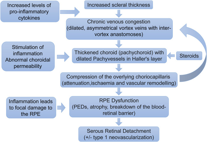
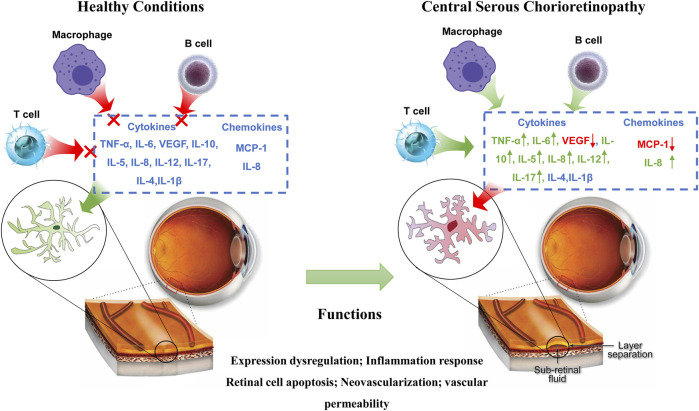
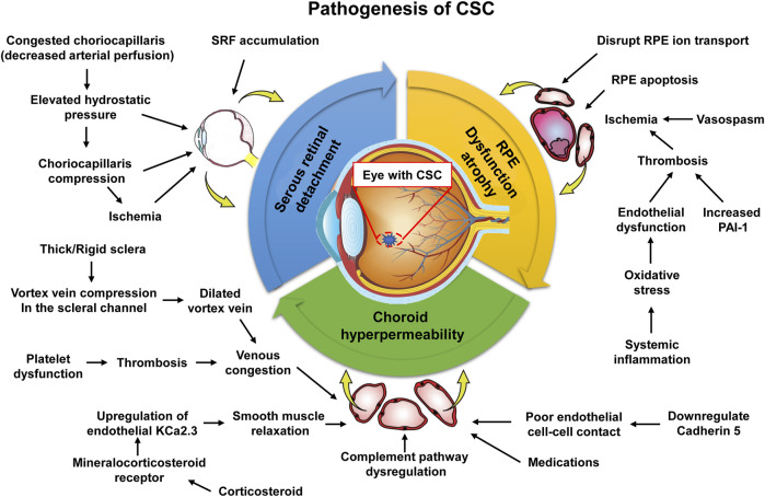
Central serous chorioretinopathy (CSC) is a leading cause of permanent vision loss, ranking fourth among macular diseases, trailing only age-related macular degeneration, diabetic retinopathy, and retinal vein obstruction. While mounting evidence implicates inflammation as a pivotal factor in the onset and advancement of CSC, the specific pathophysiological process and molecular mechanisms underlying inflammation remain incompletely understood. A complex network of cytokines, chemokines, and adhesion molecules interplay to trigger inflammatory and pathological cascades, highlighting the need for a comprehensive comprehension of the inflammation-related mechanisms behind CSC progression. In this piece, we examine the existing comprehension of CSC's pathology and pathogenesis. Additionally, we present an overview of the mechanisms underlying the onset and progression of CSC inflammation, followed by a thorough analysis and discussion of the potential of targeted inflammatory intervention for both preventing and treating CSC.
Keywords: central serous chorioretinopathy; chemokines; cytokines; inflammation; therapy.
Copyright © 2024 Shen, Kong, Wen, Wang and Huang.
Conflict of interest statement
The authors declare that the research was conducted in the absence of any commercial or financial relationships that could be construed as a potential conflict of interest.
Figures
Similar articles
-
Exosomal miR-184 in the aqueous humor of patients with central serous chorioretinopathy: a potential diagnostic and prognostic biomarker.J Nanobiotechnology. 2023 Jul 28;21(1):242. doi: 10.1186/s12951-023-02019-6. J Nanobiotechnology. 2023. PMID: 37507708 Free PMC article.
-
[Central serous chorioretinopathy].Ophthalmologe. 2021 Sep;118(9):967-980. doi: 10.1007/s00347-021-01376-7. Epub 2021 Apr 16. Ophthalmologe. 2021. PMID: 33861376 Review. German.
-
The role of inflammation in immune system of diabetic retinopathy: Molecular mechanisms, pathogenetic role and therapeutic implications.Front Immunol. 2022 Dec 13;13:1055087. doi: 10.3389/fimmu.2022.1055087. eCollection 2022. Front Immunol. 2022. PMID: 36582230 Free PMC article. Review.
-
A Review of Central Serous Chorioretinopathy: Clinical Presentation and Management.Cureus. 2022 Aug 13;14(8):e27965. doi: 10.7759/cureus.27965. eCollection 2022 Aug. Cureus. 2022. PMID: 36120212 Free PMC article. Review.
-
Risk of retinopathy in women with pregnancy-induced hypertension: a nationwide population-based cohort study of 9-year follow-up after delivery.Am J Obstet Gynecol MFM. 2023 Jul;5(7):100985. doi: 10.1016/j.ajogmf.2023.100985. Epub 2023 Apr 27. Am J Obstet Gynecol MFM. 2023. PMID: 37119970
Cited by
-
The Neutrophil to Lymphocyte Ratio and Other Full Blood Count Indices in Retinal Diseases: A Systematic Review of the Literature.Medicina (Kaunas). 2025 Jan 14;61(1):125. doi: 10.3390/medicina61010125. Medicina (Kaunas). 2025. PMID: 39859107 Free PMC article.
-
AAV2.7m8 transduction of stage 2 human retinal organoids induces highly variable responses in innate and inflammatory gene expression and cytokine secretion.Exp Eye Res. 2025 Sep;258:110478. doi: 10.1016/j.exer.2025.110478. Epub 2025 Jun 6. Exp Eye Res. 2025. PMID: 40484363
References
Publication types
LinkOut - more resources
Full Text Sources