New findings on retinal microvascular changes in patients with primary COVID-19 infection: a longitudinal study
- PMID: 38835770
- PMCID: PMC11148381
- DOI: 10.3389/fimmu.2024.1404785
New findings on retinal microvascular changes in patients with primary COVID-19 infection: a longitudinal study
Abstract
Purpose: To investigate the longitudinal alterations of retinal microvasculature in patients with primary coronavirus disease 2019 (COVID-19) infection.
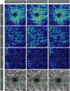
Methods: A cohort of participants, who had never been infected with COVID-19, was recruited between December 2022 and May 2023 at Peking Union Medical College Hospital in Beijing, China. Participants underwent comprehensive ophthalmologic examinations and fundus imaging, which included color fundus photography, autofluorescence photography, swept-source optical coherence tomography (SS-OCT) and SS-OCT angiography (SS-OCTA). If participants were infected with COVID-19 during the study, follow-ups with consistent imaging modality were conducted within one week and two months after recovery from the infection.
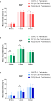
Results: 31 patients (61 eyes), with a mean age of 31.0 ± 7.2 years old, were eligible for this study. All participants contracted mild COVID-19 infection within one month of baseline data collection. The average period was 10.9 ± 2.0 days post-infection for the first follow-up and 61.0 ± 3.5 days for the second follow-up. No clinical retinal microvasculopathy features were observed during the follow-ups. However, SS-OCTA analysis showed a significant increase in macular vessel density (MVD) from 60.76 ± 2.88% at baseline to 61.59 ± 3.72%(p=0.015) at the first follow-up, which subsequently returned to the baseline level of 60.23 ± 3.33% (p=0.162) at the two-month follow-up. The foveal avascular zone (FAZ) remained stable during the follow-ups with areas of 0.339 ± 0.097mm2, 0.342 ± 0.093mm2, and 0.344 ± 0.098mm2 at the baseline, first follow-up (p=0.09) and second follow-up (p=0.052), respectively. Central macular thickness, cube volume and ganglion cell-inner plexiform layer showed a transient decrease at the first follow-up(p<0.001, p=0.039, p=0.002, respectively), and increased to baseline level at the two-month follow-up(p=0.401, p=0.368, p=0.438, respectively).
Conclusion: Mild COVID-19 infection may temporarily and reversibly impact retinal microvasculature, characterized by a transient increase in retinal blood flow during the early recovery phase, which returns to the pre-infection level two months post-infection.
Keywords: coronavirus disease 2019; foveal avascular zone; optical coherence tomography angiography; retinal microvasculature; vessel density.
Copyright © 2024 Zhang, Cheng, Chen, Yang and Chen.
Conflict of interest statement
The authors declare that the research was conducted in the absence of any commercial or financial relationships that could be construed as a potential conflict of interest.
Figures


Similar articles
-
Optical coherence tomography-based assessment of macular vessel density, retinal layer metrics and sub-foveal choroidal thickness in COVID-19 recovered patients.Indian J Ophthalmol. 2023 Feb;71(2):385-395. doi: 10.4103/ijo.IJO_1236_22. Indian J Ophthalmol. 2023. PMID: 36727324 Free PMC article.
-
[Multimodal imaging features of acute macular retinopathy in patients with COVID-19].Zhonghua Yan Ke Za Zhi. 2023 Jul 11;59(7):557-565. doi: 10.3760/cma.j.cn112142-20230109-00015. Zhonghua Yan Ke Za Zhi. 2023. PMID: 37408427 Chinese.
-
Assessment of macular microvasculature features before and after vitrectomy in the idiopathic macular epiretinal membrane using a grading system: An optical coherence tomography angiography study.Acta Ophthalmol. 2021 Nov;99(7):e1168-e1175. doi: 10.1111/aos.14753. Epub 2021 Jan 10. Acta Ophthalmol. 2021. PMID: 33423352
-
Retinal microvascular impairment in COVID-19 patients: A meta-analysis.Immun Inflamm Dis. 2022 Jun;10(6):e619. doi: 10.1002/iid3.619. Immun Inflamm Dis. 2022. PMID: 35634955 Free PMC article. Review.
-
Association between retinal vessels caliber and systemic health: A comprehensive review.Surv Ophthalmol. 2025 Mar-Apr;70(2):184-199. doi: 10.1016/j.survophthal.2024.11.009. Epub 2024 Nov 16. Surv Ophthalmol. 2025. PMID: 39557345 Review.
Cited by
-
Evaluation of Retinal and Optic Nerve Parameters in Recovered COVID-19 Patients: Potential Neurodegenerative Impact on the Ganglion Cell Layer.Diagnostics (Basel). 2025 May 9;15(10):1195. doi: 10.3390/diagnostics15101195. Diagnostics (Basel). 2025. PMID: 40428188 Free PMC article.
-
Evaluation of Retinal and Posterior Segment Vascular Changes Due to Systemic Hypoxia Using Optical Coherence Tomography Angiography.J Clin Med. 2024 Nov 7;13(22):6680. doi: 10.3390/jcm13226680. J Clin Med. 2024. PMID: 39597827 Free PMC article.
References
-
- The World Health Organization . Available online at: https://www.who.int/emergencies/diseases/novel-coronavirus-2019 (Accessed 14 June 2023).
-
- the WHO . Statement on the fifteenth meeting of the IHR (2005) Emergency Committee on the COVID-19 pandemic. Available online at: https://www.who.int/news/item/05–05-2023-statement-on-the-fifteenth-meet... (Accessed 5 May 2023).
MeSH terms
LinkOut - more resources
Full Text Sources
Medical

