. 2024 Jun 5;81(1):249.
doi: 10.1007/s00018-024-05271-7.

Legionella effectors SidC/SdcA ubiquitinate multiple small GTPases and SNARE proteins to promote phagosomal maturation

Affiliations

Legionella effectors SidC/SdcA ubiquitinate multiple small GTPases and SNARE proteins to promote phagosomal maturation

Kelong Ma et al. Cell Mol Life Sci. .

Abstract

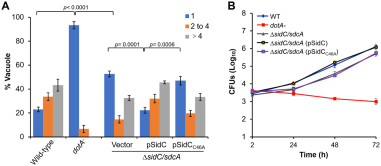
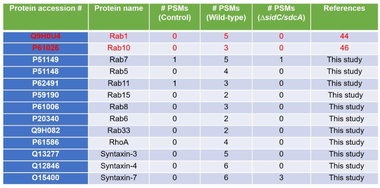
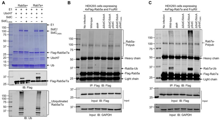
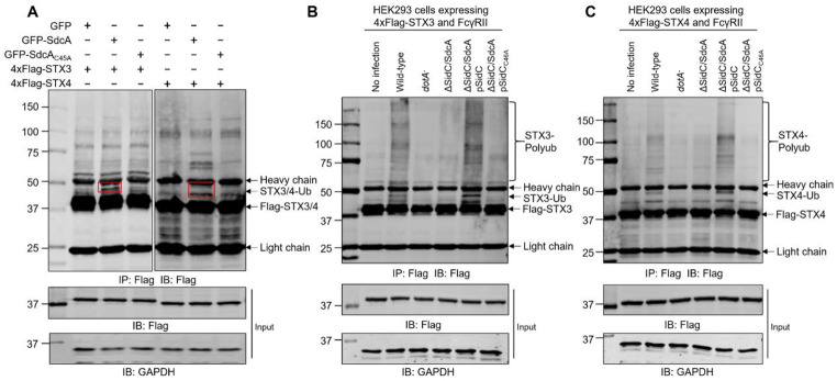
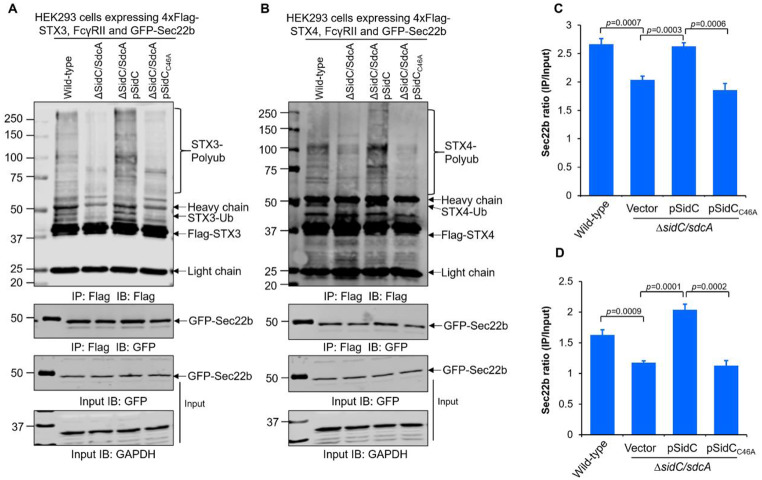
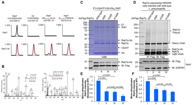
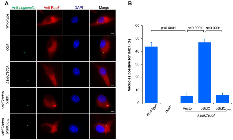
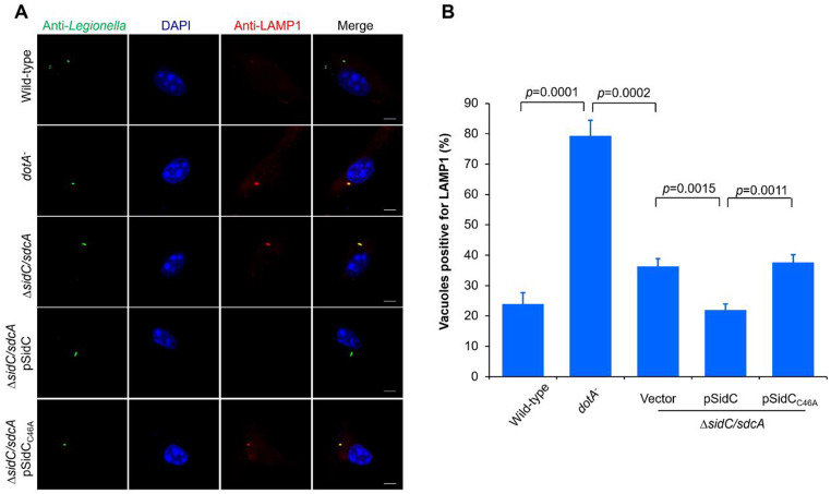
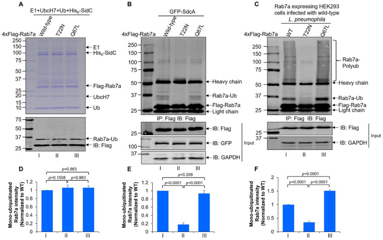
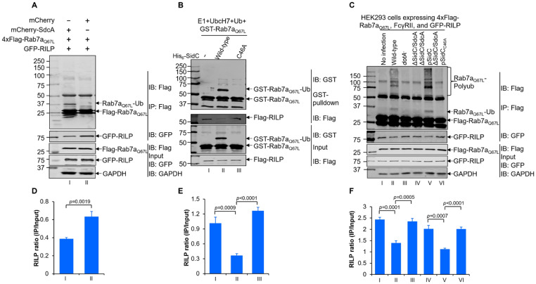
Protein ubiquitination is one of the most important posttranslational modifications (PTMs) in eukaryotes and is involved in the regulation of almost all cellular signaling pathways. The intracellular bacterial pathogen Legionella pneumophila translocates at least 26 effectors to hijack host ubiquitination signaling via distinct mechanisms. Among these effectors, SidC/SdcA are novel E3 ubiquitin ligases with the adoption of a Cys-His-Asp catalytic triad. SidC/SdcA are critical for the recruitment of endoplasmic reticulum (ER)-derived vesicles to the Legionella-containing vacuole (LCV). However, the ubiquitination targets of SidC/SdcA are largely unknown, which restricts our understanding of the mechanisms used by these effectors to hijack the vesicle trafficking pathway. Here, we demonstrated that multiple Rab small GTPases and target soluble N-ethylmaleimide-sensitive factor attachment protein receptors (SNARE) proteins are bona fide ubiquitination substrates of SidC/SdcA. SidC/SdcA-mediated ubiquitination of syntaxin 3 and syntaxin 4 promotes their unconventional pairing with the vesicle-SNARE protein Sec22b, thereby contributing to the membrane fusion of ER-derived vesicles with the phagosome. In addition, our data reveal that ubiquitination of Rab7 by SidC/SdcA is critical for its association with the LCV membrane. Rab7 ubiquitination could impair its binding with the downstream effector Rab-interacting lysosomal protein (RILP), which partially explains why LCVs avoid fusion with lysosomes despite the acquisition of Rab7. Taken together, our study reveals the biological mechanisms employed by SidC/SdcA to promote the maturation of the LCVs.

Keywords: Effector proteins; Legionella pneumophila; RILP; Rab small GTPase; SNARE proteins; Ubiquitination.


Conflict of interest statement

The authors have no relevant financial or non-financial interests to disclose.

Figures

Fig. 1
Fig. 1
The E3 ubiquitin ligase activity of SidC/SdcA is required for optimal intracellular L. pneumophila growth. Bone marrow-derived macrophages (BMDMs) were infected with wild-type, dotA, ΔsidC/sdcA, ΔsidC/sdcA (pSidC), and ΔsidC/sdcA (pSidCC46A) L. pneumophila strains at an MOI of 0.05. (A) Infected cells were fixed at 8 h postinfection and subjected to immunostaining with anti-Legionella antibodies. The number of bacteria residing in the vacuole was scored under a fluorescence microscope. One hundred phagosomes were calculated for each infection sample. (B) Intracellular growth of the bacterial strains was determined at each examined time point by counting the colony forming units (CFUs) on the plates. Data in panel A are presented as the mean ± standard deviation (SD) of three independent tests, and panel B is one representative from three independent experiments performed in triplicate
Fig. 2
Fig. 2
Candidate ubiquitination substrates of SidC/SdcA identified from L. pneumophila-infected cells by liquid chromatography-tandem mass spectrometry (LC‒MS/MS). HEK293 cells transfected with 3xHA-Ub and FcγII were either left uninfected or challenged with the wild-type or ΔsidC/sdcA L. pneumophila strain at an MOI of 20 for 2 h. The anti-HA immunoprecipitated products of the cell lysates were digested by trypsin and further analyzed by LC‒MS/MS
Fig. 3
Fig. 3
Ubiquitination of Rab5 and Rab7 by the effector protein SidC. (A) In vitro ubiquitination reactions containing E1, UbcH7, ubiquitin, 4xFlag-Rab5/Rab7, and SidC/SidCC46A were performed at 37 °C for 1 h. After termination by the addition of SDS sampling buffer, protein samples were separated by SDS‒PAGE, and Rab5/Rab7 ubiquitination was visualized by Coomassie brilliant blue (CBB) staining (upper) or Western blot analysis with anti-Flag (middle) and anti-(ubiquitin) Ub (lower) antibodies. (B-C) HEK293 cells expressing 4xFlag-Rab5/Rab7 and FcγRII were infected with the indicated L. pneumophila strains (MOI = 50). Cell lysates were prepared at 2 h postinfection with RIPA buffer, and 4xFlag-Rab5 (B) and Rab7 (C) were enriched with anti-Flag agarose and detected by Western blot using anti-Flag antibodies. The anti-glyceraldehyde-3-phosphate dehydrogenase (GAPDH) blot was included to indicate equal loading. The data shown in A, B, and C are representative of three independent experiments
Fig. 4
Fig. 4
SidC/SdcA induced ubiquitination of the t-SNARE proteins STX3 and STX4. (A) 4xFlag-STX3/STX4 was cotransfected with GFP, GFP-SdcA, or GFP-SdcAC45A into HEK293T cells. Cell lysates prepared at 24 h posttransfection were immunoprecipitated by anti-Flag agarose and analyzed by Western blot with an anti-Flag antibody. The red circle shows the modified forms of syntaxin 3 (STX3) and syntaxin 4 (STX4). (B-C) HEK293 cells transfected to produce 4xFlag-STX3/STX4 and FcγRII were either uninfected (lane 1) or infected with wild-type, dotA, ΔsidC/sdcA, ΔsidC/sdcA (pSidC), and ΔsidC/sdcA (pSidCC46A) L. pneumophila strains (lanes 2–6). At 2 h after infection, the cells were lysed, immunoprecipitated and probed with the anti-Flag antibody. The lysates probed with the antibody specific for anti-glyceraldehyde-3-phosphate dehydrogenase (GAPDH) were used as a loading control. The data shown are one representative of three independent experiments
Fig. 5
Fig. 5
Ubiquitination of syntaxin 3 (STX3) and syntaxin 4 (STX4) promotes their noncanonical pairing with the v-SNARE protein Sec22b during L. pneumophila infection. (A-B) Plasmids encoding 4xFlag-STX3/STX4, GFP-Sec22b, and FcγRII were transfected into HEK293 cells. At 24 h posttransfection, cells were then challenged with relevant L. pneumophila strains (lanes 2–5) for 2 h (MOI = 50). 4xFlag-STX3/STX4 was enriched by immunoprecipitation of the cell lysates with anti-Flag agarose, and the bead-bound proteins were further detected by Western blotting with anti-Flag and anti-GFP antibodies. The cell lysates probed with the anti-glyceraldehyde-3-phosphate dehydrogenase (GAPDH) antibody were included as a loading control. (C-D) Intensities of the GFP-Sec22b bands were quantified by ImageJ software. Noncanonical pairing between STX3 (C, related to panel A), STX4 (D, related to panel B) and Sec22b was evaluated by calculating the GFP-Sec22b ratio (IP/Input). The results shown in panels A and B are representative of three independent experiments. Values in panels C and D are the mean ± SD of three independent tests
Fig. 6
Fig. 6
SidC/SdcA ubiquitinates Rab7 at multiple lysine residues. (A) Extracted ion chromatograms of the tryptic peptides (-KKVLLK-, and -KFSNQYK-) bearing di-glycine remnants at K6 and K32 of Rab7 are shown. The unmodified reference peptide of Rab7 (-VIILGDSGVGK-) and the ubiquitin peptide (-TLSDYNIQK-) are also presented. (B) Collision-induced dissociation (CID) MS/MS spectra of Rab7 peptides carrying di-glycine remnants at K6 and K32 are shown. (C) In vitro ubiquitination of Rab7 lysine mutants by SidC. Reactions consisting of E1, UbcH7, ubiquitin, His6-SidC and each of the Rab7 derivatives were incubated at 37 °C for 30 min. After termination of the reactions by supplementation with 5x SDS sample buffer, the proteins were resolved by SDS‒PAGE and visualized by Coomassie brilliant blue (CBB) staining or Western blot using the anti-Flag antibody. (D) HEK293 cells producing wild-type Rab7, Rab7K6R, Rab7K32R, or Rab7K194R were infected with wild-type L. pneumophila for 2 h. Lysates of the infected cells were immunoprecipitated with anti-Flag beads and further analyzed by Western blotting with an antibody specific for Flag. Glyceraldehyde-3-phosphate dehydrogenase (GAPDH) was probed as the loading control. (E-F) Quantification of the mono-ubiquitinated Rab7a as shown in (C) and (D) was performed by ImageJ. Panel E is related to panel C while panel F is related to panel D. The data shown in panels C and D are representative of three independent assays. Values in panels E and F are the mean ± SD of three independent tests
Fig. 7
Fig. 7
SidC/SdcA-dependent association of Rab7 with the bacterial phagosome. BMDMs were infected with wild-type, dotA, ΔsidC/sdcA, ΔsidC/sdcA (pSidC), and ΔsidC/sdcA (pSidCC46A) L. pneumophila strains for 2 h at an MOI of 5. Fixed BMDMs were subjected to stepwise immunostaining with Legionella- (green) and Rab7- (red) specific antibodies. 2-(4-Amidinophenyl)-6-indolecarbamidine dihydrochloride (DAPI) staining was used to label the nucleus. Fluorescence signals were visualized under a Zeiss LSM880 confocal microscope. (A) Representative fluorescence micrographs of Rab7-positive and Rab7-negative LCVs in different L. pneumophila strain-challenged cells. Bar, 2 μm. (B) Percentage of Rab7-positive vacuoles. One hundred LCVs were scored for each infection sample, and values are the mean ± SD of three independent experiments
Fig. 8
Fig. 8
SidC/SdcA prevents the accumulation of LAMP1 on LCVs. BMDMs seeded on coverslips were challenged with the indicated L. pneumophila strains at an MOI of 5. Two hours after infection, the cells were fixed, permeabilized, and immunostained with anti-Legionella (green) and anti-lysosome-associated membrane proteins 1 (LAMP1) (red) antibodies. The nucleus was labeled by DAPI (blue). Immunofluorescence was observed using a Zeiss LSM880 confocal microscope. (A) Images show LAMP1 accumulation on LCVs harboring wild-type, dotA, ΔsidC/sdcA, ΔsidC/sdcA (pSidC), and ΔsidC/sdcA (pSidCC46A) L. pneumophila. Bar, 2 μm. (B) Percentage of vacuoles positively stained with LAMP1. One hundred vacuoles were calculated for each sample. Data are presented as the mean ± SD from three independent assays
Fig. 9
Fig. 9
SidC/SdcA preferred to catalyze GTP-bound active Rab7 in vivo. (A) In vitro ubiquitination of dominant-positive (Q67L) and dominant-negative (T22N) Rab7 by SidC. Reactions containing E1, UbcH7, ubiquitin, SidC and wild-type Rab7 or T22N/Q67L mutated Rab7 were performed at 37 °C for 1 h. Proteins separated by SDS‒PAGE were visualized by Coomassie brilliant blue (CBB) staining or Western blot with the anti-Flag antibody. (B) 4xFlag-tagged Rab7, Rab7T22N, or Rab7Q67L was coexpressed with GFP-SdcA in HEK293T cells. Cell lysates prepared 24 h post transfection were immunoprecipitated by anti-Flag beads. Cell lysates and the bead-enriched products were analyzed by Western blot using antibodies specific for Flag, GFP, or glyceraldehyde-3-phosphate dehydrogenase (GAPDH). (C) HEK293 cells transiently expressing FcγRII and 4xFlag-tagged Rab7, Rab7T22N, or Rab7Q67L were further infected with wild-type L. pneumophila for 2 h at an MOI of 50. 4xFlag-tagged proteins were enriched by anti-Flag immunoprecipitation and detected by Western blot using Flag-specific antibodies. The anti-glyceraldehyde-3-phosphate dehydrogenase (GAPDH) blot was probed as the loading control. (D-F) Quantification of the mono-ubiquitinated Rab7a as shown in (A), (B), and (C) was determined by ImageJ. Panel D is related to panel A, panel E is related to panel B, and panel F is related to panel C. The results from panels A, B, and C are representative of three independent experiments. Values in panels D, E, and F are the mean ± SD of three independent tests
Fig. 10
Fig. 10
Ubiquitination of Rab7 decreases its interaction with the downstream effector Rab-interacting lysosomal protein (RILP). (A) HEK293T cells were transfected with 4xFlag-Rab7Q67L, GFP-RILP, and mCherry or mCherry-SdcA. Cells were lysed and immunoprecipitated with anti-Flag agarose. The cell lysates and the bead-associated proteins were detected by Western blot using anti-Flag and anti-GFP antibodies. Glyceraldehyde-3-phosphate dehydrogenase (GAPDH) was probed as the loading control. (B) In vitro ubiquitination reactions consisting of E1, UbcH7, Ub, His6-SidC/His6-SidCC46A, and GST-Rab7Q67L were allowed to proceed for 2 h at 37 °C. Equal amount of recombinant His6-Flag-RILP was then added to each of the reaction and further incubated at 4 oC for 1 h. After enrichment with the GST magnetic beads, the interaction between GST-Rab7Q67L and His6-Flag-RILP was evaluated via Western blot using anti-GST or anti-Flag antibodies. (C) HEK293 cells transiently coexpressing 4xFlag-Rab7Q67L, FcγRII, and GFP-RILP were either uninfected (lane 2) or infected with the indicated L. pneumophila strains (lane 3–7) for 2 h (MOI = 50). After immunoprecipitation of the cell lysates by the anti-Flag beads, the association of RILP with Rab7Q67L was assessed by Western blot analysis with Flag- or GFP-specific antibodies. Glyceraldehyde-3-phosphate dehydrogenase (GAPDH) was probed as the loading control. (D-F) The intensities of GFP-RILP were determined by ImageJ software. The GFP-RILP ratio was calculated by dividing the intensities of bead-bound RILP (IP) by those in the cell lysates (Input). Panels D, E, and F are related to panels A, B, and C, respectively. The data in panels A-C are representative of three independent experiments, while those in panels D-F are the mean ± SD of three independent assays

Similar articles

Cited by

References

    1. Newton HJ, Ang DK, van Driel IR, Hartland EL. Molecular pathogenesis of infections caused by Legionella pneumophila. Clin Microbiol Rev. 2010;23:274–298. doi: 10.1128/CMR.00052-09. - DOI - PMC - PubMed
    1. Mondino S, Schmidt S, Rolando M, Escoll P, Gomez-Valero L, Buchrieser C. Legionnaires’ Disease: state of the art knowledge of Pathogenesis Mechanisms of Legionella. Annu Rev Pathol. 2020;15:439–466. doi: 10.1146/annurev-pathmechdis-012419-032742. - DOI - PubMed
    1. Isberg RR, O’Connor TJ, Heidtman M. The Legionella pneumophila replication vacuole: making a cosy niche inside host cells. Nat Rev Microbiol. 2009;7:13–24. doi: 10.1038/nrmicro1967. - DOI - PMC - PubMed
    1. Qiu J, Luo ZQ. Legionella and Coxiella effectors: strength in diversity and activity. Nat Rev Microbiol. 2017;15:591–605. doi: 10.1038/nrmicro.2017.67. - DOI - PubMed
    1. Machner MP, Isberg RR. Targeting of host rab GTPase function by the intravacuolar pathogen Legionella pneumophila. Dev Cell. 2006;11:47–56. doi: 10.1016/j.devcel.2006.05.013. - DOI - PubMed

MeSH terms

LinkOut - more resources