. 2024 Jun;23(3):e12895.
doi: 10.1111/gbb.12895.

Learning, memory and blood-brain barrier pathology in Duchenne muscular dystrophy mice lacking Dp427, or Dp427 and Dp140

Affiliations

Learning, memory and blood-brain barrier pathology in Duchenne muscular dystrophy mice lacking Dp427, or Dp427 and Dp140

Minou Verhaeg et al. Genes Brain Behav. 2024 Jun.

Abstract

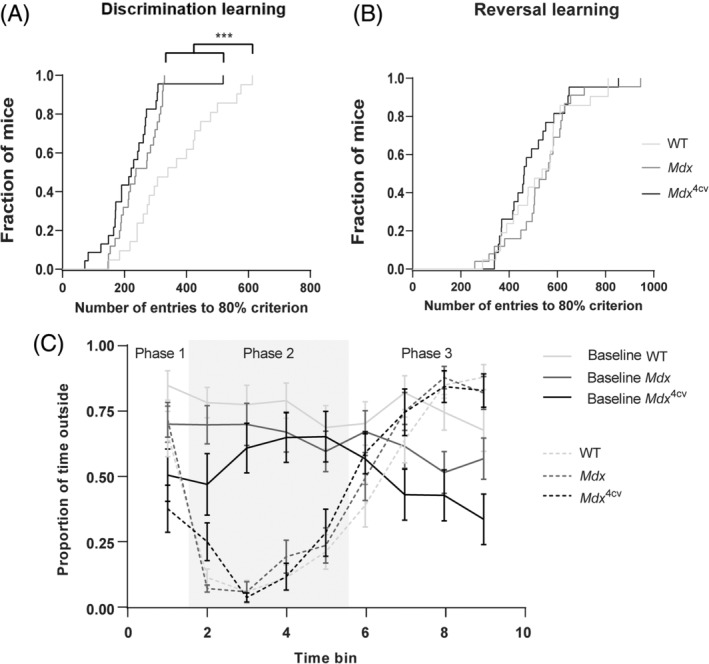
Duchenne muscular dystrophy is a severe neuromuscular disorder that is caused by mutations in the DMD gene, resulting in a disruption of dystrophin production. Next to dystrophin expression in the muscle, different isoforms of the protein are also expressed in the brain and lack of these isoforms leads to cognitive and behavioral deficits in patients. It remains unclear how the loss of the shorter dystrophin isoform Dp140 affects these processes. Using a variety of behavioral tests, we found that mdx and mdx4cv mice (which lack Dp427 or Dp427 + Dp140, respectively) exhibit similar deficits in working memory, movement patterns and blood-brain barrier integrity. Neither model showed deficits in spatial learning and memory, learning flexibility, anxiety or spontaneous behavior, nor did we observe differences in aquaporin 4 and glial fibrillary acidic protein. These results indicate that in contrast to Dp427, Dp140 does not play a crucial role in processes of learning, memory and spontaneous behavior.

Keywords: cognition; dystrophin; neuromuscular disease; spatial learning; spontaneous behavior.


Conflict of interest statement

None related to this work. For full transparency, AAR discloses being employed by LUMC, which has patents on exon skipping technology, some of which has been licensed to BioMarin and subsequently sublicensed to Sarepta. As co‐inventor of some of these patents AAR is entitled to a share of royalties. AAR further discloses being ad hoc consultant for PTC Therapeutics, Sarepta Therapeutics, Regenxbio, Alpha Anomeric, Lilly BioMarin Pharmaceuticals Inc., Eisai, Entrada, Takeda, Splicesense, Galapagos, MitoRx and Astra Zeneca. Past ad hoc consulting has occurred for: CRISPR Therapeutics, Summit PLC, Audentes Santhera, Bridge Bio, Global Guidepoint and GLG consultancy, Grunenthal, Wave and BioClinica. AAR also reports having been a member of the Duchenne Network Steering Committee (BioMarin) and being a member of the scientific advisory boards of Eisai, hybridize therapeutics, silence therapeutics, Sarepta therapeutics. Past SAB memberships: ProQR, Philae Pharmaceuticals. Remuneration for these activities is paid to LUMC. LUMC also received speaker honoraria from PTC Therapeutics, Alnylam Netherlands, Pfizer and BioMarin Pharmaceuticals and funding for contract research from Italfarmaco, Sapreme, Eisai, Galapagos, Synnaffix and Alpha Anomeric. Project funding is received from Sarepta Therapeutics and Entrada.

Figures

FIGURE 1
FIGURE 1
Overview of the experimental setup. Mice started between 10 and 15 weeks of age and underwent testing via T‐maze, Morris water maze (MWM), PhenoTyper cages and a series of MRI scans. AN, anxiety; DL, discrimination learning; RL, reversal learning; SB, spontaneous behavior.
FIGURE 2
FIGURE 2
T‐maze spontaneous alternation for WT (n = 21), mdx (n = 21) and mdx 4cv (n = 19) mice. (A) Lower levels of spontaneous alternation were observed in mdx and mdx 4cv compared with WT mice. Chance level is indicated by the dotted line. (B) Alterations per trial did not differ between strains. Asterisks indicate ***p < 0.001.
FIGURE 3
FIGURE 3
Morris water maze spatial and reversal learning for WT (n = 21), mdx (n = 25) and mdx 4cv (n = 23) mice. (A) Distance to platform average of all trials per day. All mice had the capacity to learn the platform location as indicated by the decrease in average distance required to locate the platform. Mdx mice had a decreased learning speed compared with WT and mdx 4cv mice. (B) Maximum swim velocity was significantly lower in mdx compared with WT and mdx 4cv mice. (C) Average distance swam to reach the platform zone during the probe trial. (D) Distance to platform average of all trials per day. All mice had a comparable capacity to learn the new platform location as indicated by the decrease in average distance swam to the platform. (E) Maximum swim velocity was significantly lower in mdx compared with WT and mdx 4cv mice. (F) Average distance swam to reach the platform zone during the probe trial. Asterisks indicate **p < 0.01, ***p < 0.001.
FIGURE 4
FIGURE 4
Discrimination‐ and reversal learning with cognition wall and anxiety in PhenoTyper cages for WT (n = 21), mdx (n = 23–25) and mdx 4cv (n = 22–23) mice. 80% success rate was calculated over a time window of 30 entries. (A) Survival curve of discrimination learning trials. Mdx and mdx 4cv mice were significantly faster in reaching the 80% criterium than WTs during discrimination learning. (B) Survival curves of reversal learning trials. No differences were found between groups. (C). Grey area (phase 2) represents the time the light was on. All groups showed a significant response to the light in phase 2 compared with baseline measurements of 24 h earlier. No differences were found between groups in the anxiety response. Asterisks indicate ***p < 0.001.
FIGURE 5
FIGURE 5
Spontaneous behavior in PhenoTyper cages for WT (n = 21), mdx (n = 23) and mdx 4cv (n = 22) mice. (A) Average velocity was lower in mdx mice compared with WT mice during the dark phase. (B) The duration of active behavior was shorter in both DMD models compared with WTs in the dark phase. (C) Mdx and mdx 4cv mice showed a smaller change in activity in anticipation of the start of the dark phase, compared with WT mice. (D) Long movement threshold values revealed decreases in threshold for both mdx and mdx 4cv mice compared with WTs (E) Maximum velocity of long movement segment was decreased in mdx compared with WT mice. (F) Long movement arrest threshold was significantly lower in mdx mice compared with WTs. Asterisks indicate *p < 0.05, **p < 0.01, ***p < 0.001. Cartoons are taken from Loos et al. 2014.
FIGURE 6
FIGURE 6
Blood–brain barrier permeability in WT (n = 10), mdx (n = 9) and mdx 4cv (n = 10) mice. (A) Signal intensity of the hippocampus. Mdx mice had increased T1 values compared with WTs. (B) Signal intensity of the cortex. Mdx 4cv mice showed increased T1 values compared with WTs. (C) Signal intensity of the amygdala. D) Signal intensity of the caudate putamen. Asterisks indicate **p < 0.01.
FIGURE 7
FIGURE 7
Immunohistochemical staining quantification of AQP4 and GFAP in WT (n = 5), mdx (n = 6) and mdx 4cv (n = 6) mice. (A) Representative images of AQP4 and GFAP expression in the CA1 region of the hippocampus of a WT, mdx and mdx 4cv mouse. (B) No significant differences in expression levels of AQP4 in the CA1 region of the hippocampus. (C) No significant differences in expression levels of GFAP in the CA1 region of the hippocampus. (D) Representative images of AQP4 and GFAP expression in the CA2/3 region of the hippocampus of a WT, mdx and mdx 4cv mouse. (E) No significant differences in expression levels of AQP4 in the CA2/3 region of the hippocampus. (F) No significant differences in expression levels of GFAP in the CA2/3 region of the hippocampus. (G) Representative images of AQP4 expression in the cortex of a WT, mdx and mdx 4cv mouse. (H) No significant differences in expression levels of AQP4 in the cortex. (I) Representative image of a WT mouse, showing the location of the ROIs drawn for each region.

Similar articles

Cited by

References

    1. Aartsma‐Rus A, Ginjaar IB, Bushby K. The importance of genetic diagnosis for Duchenne muscular dystrophy. J Med Genet. 2016;53:145‐151. - PMC - PubMed
    1. Emery AE. The muscular dystrophies. Lancet. 2002;359:687‐695. - PubMed
    1. Banihani R, Smile S, Yoon G, et al. Cognitive and neurobehavioral profile in boys with Duchenne muscular dystrophy. J Child Neurol. 2015;30:1472‐1482. - PubMed
    1. Hinton V, Fee R, Goldstein E, de Vivo D. Verbal and memory skills in males with Duchenne muscular dystrophy. Dev Med Child Neurol. 2007;49:123‐128. - PMC - PubMed
    1. Billard C, Gillet P, Signoret J, et al. Cognitive functions in Duchenne muscular dystrophy: a reappraisal and comparison with spinal muscular atrophy. Neuromuscul Disord. 1992;2:371‐378. - PubMed

Publication types