. 2024 Aug;11(29):e2310230.
doi: 10.1002/advs.202310230. Epub 2024 Jun 5.

Red Blood Cell Membrane-Coated Nanoparticles Enable Incompatible Blood Transfusions

Affiliations

Red Blood Cell Membrane-Coated Nanoparticles Enable Incompatible Blood Transfusions

Xuewei Yang et al. Adv Sci (Weinh). 2024 Aug.

Abstract

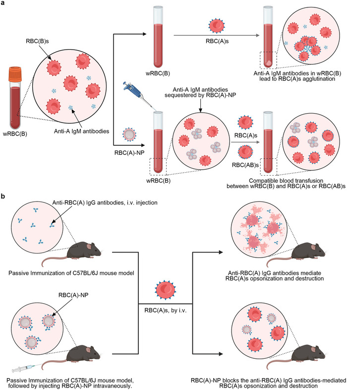
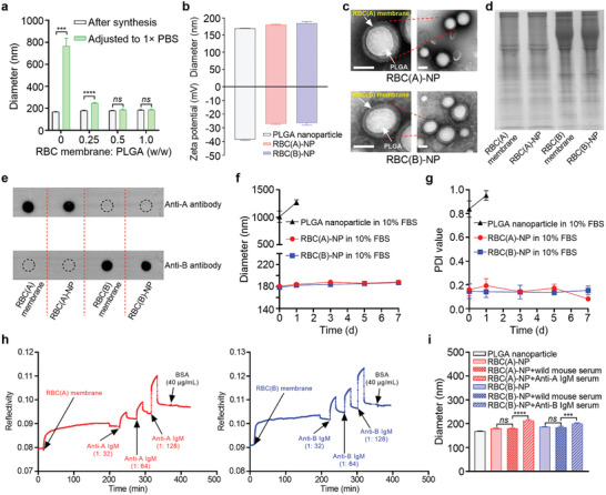
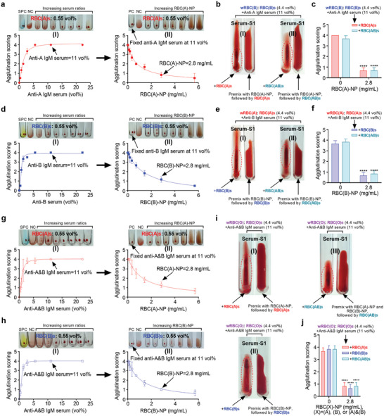
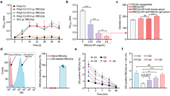
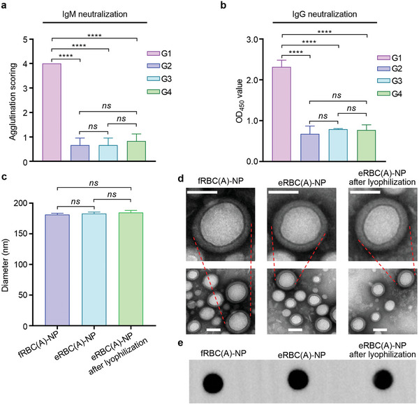
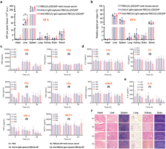
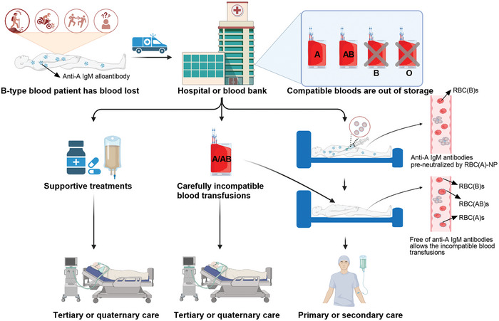
Blood transfusions save lives and improve health every day. Despite the matching of blood types being stricter than it ever has been, emergency transfusions among incompatible blood types are still inevitable in the clinic when there is a lack of acceptable blood types for recipients. Here to overcome this, a counter measure nanoplatform consisting of a polymeric core coated by a red blood cell (RBC) membrane is developed. With A-type or B-type RBC membrane camouflaging, the nanoplatform is capable of specifically capturing anti-A or anti-B IgM antibodies within B-type or A-type whole blood, thereby decreasing the corresponding IgM antibody levels and then allowing the incompatible blood transfusions. In addition to IgM, the anti-RBC IgG antibody in a passive immunization murine model can likewise be neutralized by this nanoplatform, leading to prolonged circulation time of incompatible donor RBCs. Noteworthily, nanoplatform made by expired RBCs (>42 days stored hypothermically) and then subjected to lyophilization does not impair their effect on antibody neutralization. Most importantly, antibody-captured RBC-NP do not exacerbate the risk of inflammation, complement activation, and coagulopathy in an acute hemorrhagic shock murine model. Overall, this biomimetic nanoplatform can safely neutralize the antibody to enable incompatible blood transfusion.

Keywords: antibody; biomimetic nanoparticle; cell membrane; incompatible blood transfusion; neutralization.


Conflict of interest statement

The authors declare no conflict of interest.

Figures

Scheme 1
Scheme 1
a) RBC(A)‐NP made by RBC(A)s could be utilized to neutralize anti‐A IgM antibodies in wRBC(B), leading to decreased anti‐A IgM levels, and this then allowed the transfusion of incompatible RBC(A)s or RBC(AB)s. This strategy also could be leveraged to make acceptable blood transfusions between wRBC(A) and RBC(B)s or RBC(AB)s or between wRBC(O) and RBC(A)s, RBC(B)s, or RBC(AB)s. b) Similarly, RBC(A)‐NP could be employed to block the destructive anti‐RBC(A) IgG‐induced RBC(A)s opsonization and clearance in a passive immunization murine model.
Figure 1
Figure 1
Preparation and characterization of RBC‐NP. a) Hydrodynamic sizes of RBC‐NP formulated with various RBC membranes to PLGA nanoparticles after synthesis in water and after adjusting to 1× PBS. (n = 3, mean ± SD). b) Diameters and zeta potentials of PLGA nanoparticle, RBC(A)‐NP, and RBC(B)‐NP after synthesis. RBC(A)‐NP and RBC(B)‐NP were synthesized at a weight ratio of RBC membrane to PLGA nanoparticle of 0.5: 1. (n = 3, mean ± SD). c) TEM images of RBC(A)‐NP and RBC(B)‐NP (scale bar = 100 nm). d) SDS‐PAGE protein analysis of RBC(A) membrane, RBC(A)‐NP, RBC(B) membrane, and RBC(B)‐NP at an equivalent protein concentration. All samples were imaged after Coomassie Blue staining. e) Antigen A expressed in RBC(A) membrane and RBC(A)‐NP or antigen B expressed in RBC(B) membrane and RBC(B)‐NP as determined by Dot blot. f) The colloidal stability and g) monodispersity of PLGA nanoparticle, RBC(A)‐NP, and RBC(B)‐NP in 10% FBS were monitored over 7 d. (n = 3, mean ± SD). h) The interactions between RBC(A) membrane and varying titers of anti‐A IgM serums (1: 32, 1: 64, and 1: 128 titers) or between RBC(B) membrane and varying titers of anti‐B IgM serums (1: 32, 1: 64, and 1: 128 titers), as measured by SPR. i) Diameter changes of RBC(A)‐NP or RBC(B)‐NP before and after the addition of the corresponding anti‐A (1: 128 titers) or anti‐B (1: 128 titers) IgM serums. RBC(A)‐NP or RBC(B)‐NP treated with wild mouse serums were used as controls. (n = 3, mean ± SD).
Figure 2
Figure 2
RBC‐NP blocked the corresponding IgM‐mediated incompatible blood transfusion. a) Dose‐dependent of anti‐A IgM serum (1:128 titers) agglutinated with 0.55 vol% RBC(A)s, and 11 vol% was determined to get score 4 of agglutination (I); dose‐dependent of RBC(A)‐NP against 11 vol% of anti‐A IgM serum‐mediated RBC(A)s clump, and 2.8 mg mL−1 was determined to drop the agglutination score from 4 to 1 (II). b) Images showing an experimental wRBC(B) pre‐incubated with 2.8 mg mL−1 of RBC(A)‐NP was successfully transfused with 0.55 vol% of RBC(A)s (I) or RBC(AB)s (II). The experimental wRBC(B) treated with RBC(A)s or RBC(AB)s alone were used as controls. c) Statistical analysis of agglutination scoring for (b). (n = 3, mean ± SD). d) Dose‐dependent of anti‐B IgM serum (1:128 titers) agglutinated with 0.55 vol% RBC(B)s, and 11 vol% was determined to reach score 4 of agglutination (I); dose‐dependent of RBC(B)‐NP against 11 vol% of anti‐B IgM serum‐mediated RBC(B)s aggregation, and 2.8 mg mL−1 was determined to decrease the agglutination score from 4 to 1 (II). e) Images showing an experimental wRBC(A) pre‐treated with 2.8 mg mL−1 of RBC(B)‐NP was compatibly transfused with 0.55 vol% of RBC(B)s (I) or RBC(AB)s (II). The experimental wRBC(A) treated with RBC(B)s or RBC(AB)s alone were used as controls. f) Statistical analysis of agglutination scoring for (e). (n = 3, mean ± SD). Dose‐dependent of anti‐A&B IgM serum (1:128 titers) agglutinated with 0.55 vol% g‐I) RBC(A)s or h‐I) RBC(B)s, and 11 vol% was determined to get a score 4 of agglutination; dose‐dependent of RBC(A)‐NP or RBC(B)‐NP against 11 vol% of g‐II) anti‐A&B IgM serum‐mediated RBC(A)s or h‐II) RBC(B)s clumps, and 2.8 mg mL−1 was determined to drop the agglutination score from 4 to 1 for both. i) Images showing an experimental wRBC(O) preincubated with 2.8 mg mL−1 of i‐I) RBC(A)‐NP, i‐II) RBC(B)‐NP, or i‐III) RBC(A)‐NP combined with RBC(B)‐NP were successfully transfused with 0.55 vol% of (i‐I)RBC(A)s, i‐II) RBC(B)s, and i‐III) RBC(AB)s respectively. The experimental wRBC(O) treated with RBC(A)s, RBC(B)s, or RBC(AB)s alone were used as controls. j) Statistical analysis of agglutination scoring for (i). (n = 3, mean ± SD). NC, negative control. SPC, standard positive control.
Figure 3
Figure 3
RBC‐NP protected RBCs from IgG‐mediated opsonization and clearance. a) ELISA assay detecting anti‐RBC(A) IgG antibody levels at different time points after injecting mice with various volumes of human RBC(A)s following the 100 µg of poly(I:C) prestimulation. Poly(I:C) at 100 µg only or 50 µL of RBC(A)s alone were used as controls. (n = 5, mean ± SD). b) ELISA assay detecting the various concentrations of RBC(A)‐NP for neutralizing the 10 vol% of anti‐RBC(A) IgG antibody serum. (n = 3, mean ± SD). c) Diameter changes of RBC(A)‐NP before and after incubation of anti‐RBC(A) IgG antibody serum. RBC(A)‐NP treated with wild mouse serum was used as a control. (n = 3, mean ± SD). d) Flow cytometry picture of DiD‐labeled RBC(A)s (left). Statistical analysis of labeling efficiency (right). Naïve RBC(A)s sample was employed as the negative control. (n = 3, mean ± SD). e) The remaining ratios of DiD‐labeled RBC(A)s at planned time points following the injection of various doses of RBC(A)‐NP into an established anti‐RBC(A) IgG antibody passive murine model. (n = 4, mean ± SD). f) The remaining ratio of DiD‐labeled RBC(A)s at the first 1 min following the injections of various doses of RBC(A)‐NP into an established anti‐RBC(A) IgG antibody passive murine model. (n = 4, mean ± SD). In e,f), five groups were below, G1: mice treated with PBS followed by injection of DiD‐labeled RBC(A)s; G2: mice treated with anti‐RBC(A) IgG serum followed by injection of DiD‐labeled RBC(A)s; G3: mice treated with anti‐RBC(A) IgG serum, followed by 6.3 mg kg−1 of RBC(A)‐NP and injection of DiD‐labeled RBC(A)s; G4: mice treated with anti‐RBC(A) IgG serum, followed by 25 mg kg−1 of RBC(A)‐NP and injection of DiD‐labeled RBC(A)s; G5: mice treated with anti‐RBC(A) IgG serum, followed by 100 mg kg−1 of RBC(A)‐NP and injection of DiD‐labeled RBC(A)s.
Figure 4
Figure 4
Assessment of eRBC‐NP functionality before and after lyophilization/resuspension. a) Protective effect of various RBC membrane‐based formulations against anti‐A IgM antibody‐mediated agglutination on RBC(A)s. (n = 3, mean ± SD). G1: anti‐A IgM serum only; G2: fRBC(A)‐NP pre‐incubated with anti‐A IgM serum; G3: eRBC(A)‐NP pre‐incubated with anti‐A IgM serum; G4: eRBC(A)‐NP following lyophilization and resuspension was pre‐incubated with anti‐A IgM serum. b) Neutralization effect of various RBC membrane‐based formulations against anti‐RBC(A) IgG serum. (n = 3, mean ± SD). G1: anti‐RBC(A) IgG serum alone; G2: fRBC(A)‐NP pre‐incubated with anti‐RBC(A) IgG serum; G3: eRBC(A)‐NP pre‐incubated with anti‐RBC(A) IgG serum; G4: eRBC(A)‐NP following lyophilization and resuspension was pre‐incubated with anti‐RBC(A) IgG serum. c) Hydrodynamic sizes of fRBC(A)‐NP, eRBC(A)‐NP, and eRBC(A)‐NP after treatment of lyophilization and resuspension. (n = 3, mean ± SD). d) Morphological images of fRBC(A)‐NP, eRBC(A)‐NP, and eRBC(A)‐NP as visualized by TEM. Bar = 50 nm. e) Antigen A profiles expressed in fRBC(A)‐NP, eRBC(A)‐NP, and eRBC(A)‐NP after treatment of lyophilization and resuspension were tested by Dot blot.
Figure 5
Figure 5
Biodistribution and biocompatibility of IgM or IgG‐sequestered RBC‐NP in a volume‐targeted hemorrhagic shock mouse model. a) Fluorescence intensity per gram of tissue or b) relative signal per organ for dissected major organs from mice 24 h after intravenous given of anti‐A IgM or anti‐RBC(A) IgG‐sequestered RBC(A)‐(DiD)NP into the hemorrhagic shock mice. Mice treated with PBS or RBC(A)‐(DiD)NP complexed with wild mouse serum were used as controls. (n = 3, mean ± SD). The serum concentrations regarding coagulation‐related factors including c) D2D and Fbg, complement components including d) C1q, C3, C3a, C5a, and sC5b‐9, allergic toxins including d) C3a and C5a, and inflammatory cytokines including e) IL‐6, TNF‐α, and MCP‐1, were measured by the corresponding ELISA kits on 2, 8, and 24 h post‐injection of four different treatments in the hemorrhagic shock mice. f) H&E staining of histology sections from major organs 24 h after different treatments. Scale bar = 50 µm. From c–f), four different treatments, including the hemorrhagic shock mice treated with RBC(A)‐NP complexed with anti‐A IgM or anti‐RBC(A) IgG, or RBC(A)‐NP pretreated with wild mouse serum or PBS only. (n = 5, mean ± SD).
Figure 6
Figure 6
RBC‐NP enables incompatible blood transfusions in an emergency. By neutralization of anti‐A IgM antibodies in B‐type blood recipients in an emergency where there is a lack of compatible RBC resources in hospitals or blood banks, RBC(A)‐NP enables the incompatible transfusion of donor RBCs, including RBC(A)s or RBC(AB)s. Also, this strategy could be employed to compatibly transfuse RBC(B)s or RBC(AB)s into A‐type blood recipients or transfuse RBC(A)s, RBC(B)s, or RBC(AB)s into O‐type blood recipients when incompatible blood transfusion‐related IgM antibodies could be first neutralized by the corresponding RBC‐NP. Similarly, RBC‐NP could be leveraged to block the destructive IgG‐induced RBC opsonization and clearance. This strategy can save lives or improve health in emergencies; otherwise, patients might be subjected to tertiary or quaternary care after supportive treatments or carefully incompatible blood transfusions. Overall, this strategy is expected to be applied in different blood group systems, including ABO, Rh, and others in emergencies, and lessens the tight blood supply worldwide.

Similar articles

Cited by

References

    1. D'Alessandro A., Liumbruno G., Blood Transfus. 2019, 17, 255. - PMC - PubMed
    1. Yu X., Wang Z., Shen Y., Liu Z., Wang H., Zhang S., Gan J., Xue F., Han W., Shi X., Hu Y., Wang L., Li N., Wu P., Yang C., Jiang J., Bull. World Health Organ. 2020, 98, 10. - PMC - PubMed
    1. Fahimnia B., Jabbarzadeh A., Ghavamifar A., Bell M., Int. J. Prod. Econ. 2017, 183, 700.
    1. Gniadek T. J., Mallek J., Wright G., Saporito C., AbiMansour N., Tangazi W., Rogers G., Zahara Z., Cummings G., Kaul K., Kang J., Transfusion 2020, 60, 1470. - PMC - PubMed
    1. Yang J., Cai N., Zhai H., Zhang J., Zhu Y., Zhang L., Sci. Rep. 2016, 6, 37458. - PMC - PubMed

LinkOut - more resources