. 2024 Jun 25;4(6):1566-1580.
doi: 10.1158/2767-9764.CRC-24-0049.

Activation of the Mevalonate Pathway in Response to Anti-cancer Treatments Drives Glioblastoma Recurrences Through Activation of Rac-1

Affiliations

Activation of the Mevalonate Pathway in Response to Anti-cancer Treatments Drives Glioblastoma Recurrences Through Activation of Rac-1

Ling He et al. Cancer Res Commun. .

Abstract

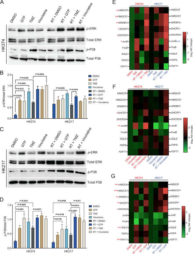
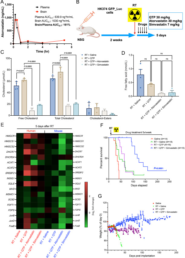
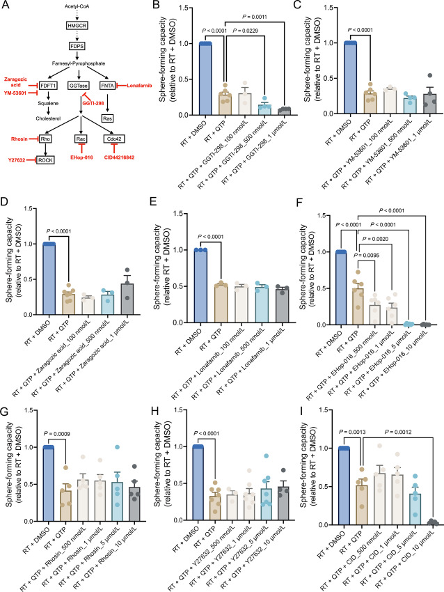
Glioblastoma (GBM) is the deadliest adult brain cancer. Under the current standard of care, almost all patients succumb to the disease and novel treatments are urgently needed. Recognizing that GBMs are addicted to cholesterol, past clinical trials have repurposed statins against GBM but failed. The purpose of this study was to test whether treatments that upregulate the cholesterol biosynthesis pathway in GBM would generate a metabolic vulnerability that can be exploited using statins and to determine the underlying mechanisms.Effects of radiotherapy and temozolomide or dopamine receptor antagonists on the mevalonate pathway in GBM were assessed in vitro and in vivo. The impact of statins on self-renewal of glioma stem cells and median survival was studied. Branches of the mevalonate pathway were probed to identify relevant effector proteins.Cells surviving combination treatments that converge in activating the immediate early response, universally upregulated the mevalonate pathway and increased stemness of GBM cells through activation of the Rho-GTPase Rac-1. Activation of the mevalonate pathway and Rac-1 was inhibited by statins, which led to improved survival in mouse models of glioblastoma when combined with radiation and drugs that target the glioma stem cell pool and plasticity of glioma cells.We conclude that a combination of dopamine receptor antagonists and statins could potentially improve radiotherapy outcome and warrants further investigation.

Significance: Combination therapies that activate the mevalonate pathway in GBM cells after sublethal treatment enhance self-renewal and migratory capacity through Rac-1 activation, which creates a metabolic vulnerability that can be further potentially exploited using statins.


Figures

FIGURE 1
FIGURE 1
Combination therapies that converge on the immediate-early response to radiation through the MAPK cascade universally upregulate the mevalonate pathway. A and C, Western blotting of p-ERK, total ERK, p-P38, and total P38 in patient-derived GBM HK374 and HK217 cell lines upon radiation (RT) in combination with QTP (10 µmol/L) or TMZ (1 mmol/L) or vincristine (250 nmol/L) at 2 hours after treatment. B and D, The densitometry measurements of p-ERK/total ERK and p-P38/total P38 using Image J. E, Heat map showing the results of qRT-PCR for the cholesterol biosynthesis–related genes in both HK374 and HK217 cells treated with radiation in the presence or absence of TMZ (1 mmol/L) for 2 consecutive days. F, Heat map showing the results of qRT-PCR for the cholesterol biosynthesis–related genes in both HK374 and HK217 cells treated with radiation in the presence or absence of vincristine (250 nmol/L) for 24 hours. G, Heat map showing the results of qRT-PCR for the cholesterol biosynthesis–related genes in both HK374 and HK217 cells treated with radiation in the presence or absence of QTP (10 µmol/L) for 2 consecutive days. All experiments have been performed with at least three biological independent repeats. P-values were calculated using one-way ANOVA. The P-values listed in the heat maps were from the comparison of RT + DMSO with RT + TMZ (E) or RT + Vincristine (F) or RT + QTP (G). *, P < 0.05; **, P < 0.01; ***, P < 0.001.
FIGURE 2
FIGURE 2
Upregulation of the mevalonate pathway in response to anti-cancer treatments in vivo is restricted to glioma cells. A, Schematic of the experimental design underlying Fig. 2. B–F, qRT-PCR for the cholesterol biosynthesis–related genes in the tumor specimen harvested from the PDOX GBM mouse model at day 2 after the treatment of radiation in combination with QTP (30 mg/kg) or TMZ (50 mg/kg) or ONC201 (50 mg/kg) or solvent control using the human-specific primers. G–K, qRT-PCR for the cholesterol biosynthesis–related genes in the tumor specimen harvested from the PDOX GBM mouse model at day 2 after the treatments using the mouse-specific primers. L, The levels of total, free cholesterol and cholesterol-esters in the tumors harvested from the PDOX GBM mouse model at day 2 after the treatments. M, The concentration of free fatty acid in the tumors harvested from the PDOX GBM mouse model at day 2 after the treatments. All experiments have been performed with at least three biological independent repeats. P-values were calculated using one-way ANOVA. ns: not significant.
FIGURE 3
FIGURE 3
Statins reduce treatment-induced upregulation of cholesterol biosynthesis in PDOXs. A, Brain and plasma levels of atorvastatin in C57BL/6 mice after a single injection (atorvastatin – 30 mg/kg, i.p.). B, Schematic of the experimental design underlying Fig. 3. C, The levels of total, free cholesterol and cholesterol-esters in the tumors harvested from the PDOX GBM mouse model at day 5 after the treatment of radiation in combination with QTP (30 mg/kg), atorvastatin (30 mg/kg) or simvastatin (7 mg/kg). D, The concentration of free fatty acid in the tumors harvested from the PDOX GBM mouse model at day 5 after the treatments. E, Heat map showing the results of qRT-PCR for the cholesterol biosynthesis–related genes in the tumor specimen harvested from the PDOX GBM mouse model at day 5 after the treatments using the human- and mouse-specific primers. F, Survival curves for NSG mice implanted intracranially with 3 × 10⁵ HK374-GFP-Luciferase glioma cells and grafted for 3 days. Mice were irradiated and treated with saline or QTP (30 mg/kg, s.c., 5-day on/2-day off schedule) or triple combination of radiation plus QTP and simvastatin (7 mg/kg, i.p., 5-day on/2-day off schedule) continuously until they reached the study endpoint. log-rank (Mantel–Cox) test for comparison of Kaplan–Meier survival curves. G, Weight curves for the NSG mice in different treatment groups. All experiments have been performed with at least three biological independent repeats. P-values were calculated using one-way ANOVA. ns: not significant.
FIGURE 4
FIGURE 4
Statins improve median survival in mouse models of GBM undergoing fractionated irradiation. A, Heat map showing the results of qRT-PCR for the cholesterol biosynthesis–related genes in HK374 cells treated with radiation in the presence or absence of ONC201 (a single treatment of 2.5 µmol/L) at 48 and 72 hours. P-values were calculated using unpaired Student t test for A. *, P < 0.05. B, Survival curves for C57BL/6 mice implanted intracranially with 2 × 10⁵ GL261-GFP-Luciferase mouse glioma cells and grafted for 7 days. Mice were irradiated and weekly treated with saline or ONC201 (50 mg/kg, i.p.) or triple combination of radiation plus ONC201 (weekly) and atorvastatin (30 mg/kg, i.p., 5-day on/2-day off schedule) continuously until they reached the study endpoint. log-rank (Mantel–Cox) test for comparison of Kaplan–Meier survival curves. C, H&E-stained coronal sections of the C57BL/6 mice brains implanted with GL261-GFP-Luc cells which were irradiated and treated continuously with ONC201 in the presence or absence of atorvastatin until they met the criteria for study endpoint. D, Schematic of the experimental design of fractionated irradiation in syngeneic mouse model of GBM. E, Kaplan–Meier survival curves for C57BL/6 mice implanted intracranially with GL261-GFP-Luciferase mouse glioma cells and treated with either a single fraction of 0 or 10 Gy or five daily fractions of 3 Gy each and daily doses of either saline, QTP (30 mg/kg, s.c.), or QTP plus atorvastatin (30 mg/kg, i.p.). After completion of the radiation treatment all animals were treated with QTP plus atorvastatin until they reached criteria for euthanasia. log-rank (Mantel–Cox) test for comparison of Kaplan–Meier survival curves. F, H&E-stained coronal sections of the C57BL/6 mice brains from the groups of 5 × 3 Gy → QTP + atorvastatin, 5 × 3 Gy + QTP → QTP + atorvastatin and 5 × 3 Gy + QTP + atorvastatin → QTP + atorvastatin.
FIGURE 5
FIGURE 5
Identification of Rac1 as the potential contributor for maintaining the stemness of surviving glioma cells. A, Cholesterol biosynthesis pathway and the key selected inhibitors. B–E, Sphere-forming capacity of HK374 spheres treated either with GGTI-298 (GGTase inhibitor) or YM-53601 (squalene synthase inhibitor) or Zaragozic acid (squalene synthase inhibitor) or Lonafarnib (farnesyltransferase inhibitor) at 100, 500 nmol/L, 1 µmol/L concentrations when combined with radiation and QTP (10 µmol/L). F–I, Sphere-forming capacity of HK374 spheres treated either with Ehop-016 (Rac GTPase inhibitor) or Rhosin (RhoA-specific inhibitor) or Y27632 (ROCK1/2 inhibitor) or CID44216842 (Cdc42-selective inhibitor) at 500 nmol/L, 1, 5, 10 µmol/L concentrations when combined with radiation and QTP. All experiments have been performed with at least three biological independent repeats. P-values were calculated using one-way ANOVA.
FIGURE 6
FIGURE 6
Treatment-induced upregulation of the mevalonate pathway in glioma affects stemness through prenylation of Rac1. A, HK374 cells were treated with radiation in the presence or absence of QTP (10 µmol/L) and/or atorvastatin (1 µmol/L) for 48 hours. The activated Rac1 was immunoprecipitated by 10 µg PAK-PBD agarose beads from the whole cell lysates and subjected to immunoblotting against Rac1, along with the total proteins. His-tagged Rac1 protein serves as the positive control. B, The densitometry measurements of activated Rac1/total Rac1 using Image J. C, Transwell migration assay of HK374 cells pretreated with radiation in the presence or absence of QTP (10 µmol/L) and/or atorvastatin (1 µmol/L) for 48 hours. D, The quantification of migrated cells using Image J. E, Confocal images of microtubules in HK374 cells treated with radiation in the presence or absence of QTP (10 µmol/L) and/or atorvastatin (1 µmol/L) for 48 hours. White arrowheads: filopodia. Yellow arrowheads: TNTs. F and G, Rac1 knockdown efficiency was evaluated at both mRNA (qRT-PCR) and protein (Western blotting) levels at day 2 and day 7 after siRNA transfection. β-actin was used as the loading control and the densitometry measurements of Rac1 were performed using Image J. H and I, Clonogenic assay of siCtrl or siRac1 transfected HK374 cells treated with radiation in the presence or absence of QTP (10 µmol/L) and/or atorvastatin (1 µmol/L) for 7 days and the resulting plating efficiencies. J, Sphere-forming capacity of siCtrl or siRac1 HK374 spheres treated with radiation in the presence or absence of QTP (10 µmol/L) and/or atorvastatin (1 µmol/L). K, log fraction plot generated by extreme limiting dilution assay (ELDA), where the y axis “log fraction nonresponding” indicates frequency of cells incapable of forming clonal spheres and the x axis “dose (number of cells)” indicates number of cells per mL. The slope of the line is the log-active cell fraction, and dotted lines give the 95% confidence interval. All experiments have been performed with at least three biological independent repeats. P-values were calculated using one-way ANOVA for B and D, unpaired Student t tests for F and G; two-way ANOVA for I and J. ns: not significant.

Update of

Similar articles

Cited by

References

    1. Eramo A, Ricci-Vitiani L, Zeuner A, Pallini R, Lotti F, Sette G, et al. . Chemotherapy resistance of glioblastoma stem cells. Cell Death Differ 2006;13:1238–41. - PubMed
    1. Bao S. Glioma stem cells promote radioresistance by preferential activation of the DNA damage response. Nature 2006;444:756–60. - PubMed
    1. Bhat K, Saki M, Vlashi E, Cheng F, Duhachek-Muggy S, Alli C, et al. . The dopamine receptor antagonist trifluoperazine prevents phenotype conversion and improves survival in mouse models of glioblastoma. Proc Natl Acad Sci U S A 2020;117:11085–96. - PMC - PubMed
    1. Bhat K, Saki M, Cheng F, He L, Zhang L, Ioannidis A, et al. . Dopamine receptor antagonists, radiation, and cholesterol biosynthesis in mouse models of glioblastoma. J Natl Cancer Inst 2021;113:1094–104. - PMC - PubMed
    1. He L, Bhat K, Ioannidis A, Zhang L, Nguyen NT, Allen JE, et al. . Effects of the DRD2/3 antagonist ONC201 and radiation in glioblastoma. Radiother Oncol 2021;161:140–7. - PMC - PubMed

Publication types

MeSH terms