. 2024 Jun 11;121(24):e2320867121.
doi: 10.1073/pnas.2320867121. Epub 2024 Jun 5.

OGA mutant aberrantly hydrolyzes O-GlcNAc modification from PDLIM7 to modulate p53 and cytoskeleton in promoting cancer cell malignancy

Affiliations

OGA mutant aberrantly hydrolyzes O-GlcNAc modification from PDLIM7 to modulate p53 and cytoskeleton in promoting cancer cell malignancy

Chia-Wei Hu et al. Proc Natl Acad Sci U S A. .

Abstract

O-GlcNAcase (OGA) is the only human enzyme that catalyzes the hydrolysis (deglycosylation) of O-linked beta-N-acetylglucosaminylation (O-GlcNAcylation) from numerous protein substrates. OGA has broad implications in many challenging diseases including cancer. However, its role in cell malignancy remains mostly unclear. Here, we report that a cancer-derived point mutation on the OGA's noncatalytic stalk domain aberrantly modulates OGA interactome and substrate deglycosylation toward a specific set of proteins. Interestingly, our quantitative proteomic studies uncovered that the OGA stalk domain mutant preferentially deglycosylated protein substrates with +2 proline in the sequence relative to the O-GlcNAcylation site. One of the most dysregulated substrates is PDZ and LIM domain protein 7 (PDLIM7), which is associated with the tumor suppressor p53. We found that the aberrantly deglycosylated PDLIM7 suppressed p53 gene expression and accelerated p53 protein degradation by promoting the complex formation with E3 ubiquitin ligase MDM2. Moreover, deglycosylated PDLIM7 significantly up-regulated the actin-rich membrane protrusions on the cell surface, augmenting the cancer cell motility and aggressiveness. These findings revealed an important but previously unappreciated role of OGA's stalk domain in protein substrate recognition and functional modulation during malignant cell progression.

Keywords: O-GlcNAcase; O-GlcNAcylation; PDLIM7; cancer; deglycosylation.


Conflict of interest statement

Competing interests statement:The authors declare no competing interest.

Figures
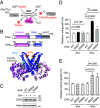
Fig. 1.
Fig. 1.
Cancer-derived point mutation S652F on OGA’s stalk domain promoted aberrant cell growth. (A) Reversible O-GlcNAcylation is catalyzed by O-GlcNAc transferase (OGT) and O-GlcNAcase (OGA). (B, Top): Schematic of OGA and truncated OGAcryst domain structures. The catalytic and stalk domains are shown in pink and blue, respectively. Intrinsically disordered regions are shown in white. pHAT, pseudo histone acetyltransferase domain. Bottom: The crystal structure of dimeric OGAcryst–D175N in complex with an O-GlcNAcylated peptide substrate (PDB: 5VVU) (22). The coloring is the same as in the schematic (Top). (C) Western blot showing the doxycycline (dox)-induced expression of WT OGA and stalk domain mutants in T-REx-293 cells (24 h dox treatment). (D) Relative growth of T-REx-293 cells expressing WT OGA or stalk domain mutant (40 h dox induction) detected by resazurin assay (n = 4). (E) Relative quantitation of the colony growth from soft agar assay using T-REx-293 cells with dox-induced expression of Flag-OGA (WT or mutant) (n = 3). Representative examples of colony growth are shown in SI Appendix, Fig. S1B. Quantified data are presented as mean ± SD. *P < 0.05; ***P < 0.001. P-values were determined by one-tailed student’s unpaired (D) or paired (E) t test.
Fig. 2.
Fig. 2.
Quantitative proteomic analyses of OGA stalk domain mutants revealed protein network perturbations. (A) Flow-chart of the integrative proteomic analyses of T-REx-293 cells with dox-induced expression of OGA (WT or mutant). IP, immunoprecipitation. (BD) Volcano plot and quantitation of the proteome (B), OGA interactome (C), and O-GlcNAcome (D). (E) The semiconsensus sequences of OGA S652F down-regulated, OGA R586A up-regulated, and total identified O-GlcNAcylation sites from O-GlcNAcome analyses. Residues depicted with the residue height over the statistical significance threshold (P < 0.05) were considered position-specifically enriched. (F) The major protein association networks and their representative functions dysregulated by OGA-S652F. Proteins identified from different proteomes were shown as indicated nodes. The light-yellow background highlights the subnetwork associated with p53. The pink arrow indicates PDLIM7.
Fig. 3.
Fig. 3.
OGA stalk domain mutant S652F dysregulated the O-GlcNAc hydrolysis from PDLIM7, a regulator of p53 and cell malignancy. (A) Western blot of the immunoprecipitated cMyc–PDLIM7 complexes from T-REx-293 cells with dox-inducible Flag-OGA (WT/S652F). Cells were transfected with cMyc–PDLIM7 for 24 h followed by 27 h dox treatment. O-GlcNAcylation was detected with CTD110.6 antibody. IP, immunoprecipitation. (B) Western blot of the recombinantly purified cMyc–PDLIM7 and His-OGA from in vitro OGA deglycosylation reactions. TMG, thiamet-G. (C) Western blot of the T-REx-293 cells with 24 h dox induction of shPDLIM7-3′UTR. Cycloheximide (CHX) was added to the cells 6 h prior to cell harvesting. Dox, doxycycline. (D, Top) The wound healing of H460 cells with dox induction of shPDLIM7-3′UTR. Bottom, western blot of the nuclear extracts from the same dox-treated cells. The dox concentration used for the wound healing assay was bolded. (E) Relative quantitation of the wound healing efficiency from (D) (n = 3). All quantified data are presented as mean ± SD. *P < 0.05. P-values were determined by one-tailed student’s unpaired t test. (F, Top) The EThcD MS/MS spectrum of PDLIM7 peptide containing the O-GlcNAcylated S89 residue. Bottom, chemical structure of the hypothetical fragmentation of biotin-conjugated oxonium ion.
Fig. 4.
Fig. 4.
PDLIM7 regulated the cellular abundance of p53 at both protein and transcription levels in an O-GlcNAc-dependent manner. (A) Western blot of the immunoprecipitated cMyc–PDLIM7 (WT/S89A) from HEK293 cells with 48 h of transient expression. O-GlcNAcylation was detected by CTD110.6 antibody. IP, immunoprecipitation. (B) Western blot of different cancer cells transiently expressed cMyc–PDLIM7 (WT/S89A) and HA–p53 for 48 h. For H1299 and HeLa cells, p53 was detected using nuclear extracts due to the low protein abundance. (C) Western blot of H1299 cells stably expressing cMyc–PDLIM7 (WT/S89A) with 24 h dox induction of shPDLIM7-3′UTR. Samples were prepared similarly as in described in (B). (D) Western blot of T-REx-293 (Left) and H460 (Right) cells with dox-inducible shPDLIM7 -3′UTR. Cells were transfected with cMyc–PDLIM7 (WT/S89A) and HA–p53 for 24 h followed by 24 h dox treatment. CHX was added at the indicated time points prior to cell harvesting. Red arrows highlight the p53 bands with significant change between WT and S89A cells. CHX, cycloheximide. (E) Western blot of MG132 (MG)-treated H1299 cells with dox-inducible cMyc–PDLIM7 (WT/S89A) and shPDLIM7-3′UTR. Cell transfection and induction were prepared similarly as described in (D) prior to MG treatment. Red arrows highlight the p53 bands in S89A cells (+MG/−MG). (F) The relative protein levels of HA–p53 (+MG/−MG) in WT and S89A cells quantified from (E) (n = 4). (G) Left, real-time qPCR detected the relative level of p53 transcripts from T-REx-293 cells with 48 h dox induction of cMyc–PDLIM7 (WT/S89A) and shPDLIM7-3′UTR. Data were quantified from four independent analyses with four technical replicates each. Right, western blot of the same cell culture used for real-time qPCR (Left). Dox, doxycycline. Quantified data are presented as mean ± SD. *P < 0.05. P-values were determined by one-tailed student’s paired t test.
Fig. 5.
Fig. 5.
Aberrant deglycosylation of PDLIM7 by OGA stalk domain mutant S652F modulated p53 ubiquitination primarily through MDM2 in cells. (A) Western blot of the immunoprecipitated HA–p53 complexes from HEK293 cells. Transient expression of cMyc–PDLIM7 (WT/S89A), HA–p53, and ubiquitin (Ub) was performed for 45 h prior to MG132 (MG) treatment. IP, immunoprecipitation. (B, Top) Relative quantitation of Ub–p53 and its bound MDM2 in a (n = 3). Bottom, relative quantitation of Ub-PDLIM7 and its bound MDM2 in (C) (n = 4). (C) Western blot of the immunoprecipitated cMyc–PDLIM7 (WT/S89A) complexes from T-REx-293 cells with dox-inducible shPDLIM7-3’UTR. Cells were prepared similarly as described in (A). Cell were treated with dox 24 h posttransfection. (D) Western blot of the immunoprecipitated HA–p53 complexes from HEK293 cells treated with Nutlin-3a (Nut). Cells were transiently expressed with cMyc–PDLIM7 (WT/S89A), HA–p53, and Ub for 48 h followed by 12 h of Nut and 3 h of MG treatments. (EG) Relative quantitation (+Nut/−Nut) of p53 bound MDM2 (E), Ub–p53 (F), and p53 bound PDLIM7 (WT/S89A) (G) from (D) (n = 3). (H) Western blot of the immunoprecipitated cMyc–PDLIM7 complexes from Nut-treated T-REx-293 cells with dox-inducible shPDLIM7-3′UTR. Cells were prepared similarly as described in (C and D). (IK) Relative quantitation (+Nut/−Nut) of PDLIM7-bound MDM2 (I), Ub-PDLIM7 (J), and PDLIM7 (WT/S89A) bound p53 (K) from (H) (n = 3). (L) Western blot of reciprocal IP of HA–p53 or cMyc–PDLIM7 complexes from T-REx-293 cells with dox-inducible Flag-OGA (WT/S652F) and shOGA-3′UTR. Cells were prepared similarly as described in (C). (M, Top) Soft agar assay of T-REx-293 cells with 48 h dox induction of Flag-OGA (WT/S652F) and shOGA-3′UTR. Bottom, Western blot of the same cell culture used in soft agar assay (Top). (N) Relative quantitation of the colony growth from (M) (n = 3). Quantified data are presented as mean ± SD. *P < 0.05; **P < 0.01. P-values were determined by one-tailed student’s paired t test.
Fig. 6.
Fig. 6.
The O-GlcNAc status of PDLIM7 modulated the density of actin protrusions and malignant cell invasion. (A) Left and Middle, wound healing assay of H1299 cells (±HA–p53) with dox-inducible cMyc–PDLIM7 (WT/S89A) and shPDLIM7-3′UTR. Right, Western blot detecting the expression of PDLIM7 from the same cells. (B) Representative images of p53-stably expressing H1299 cells with dox-inducible shPDLIM7-3′UTR cells ± cMyc–PDLIM7 (WT/S89A) stained with F-actin (green), C-Myc PDLIM7 (red), and nucleus (blue). Cells were dox-treated for 48 h. White arrows indicate the representative filopodia or filopodia-like protrusions. (Scale bar, 15 μm.) Two additional datasets are shown in SI Appendix, Fig. S7 A and B. (C) Scatter plot of the quantified filopodia density (total length of filopodia/total length of the corresponding cell edge) from (B) (WT, n = 72; S89A, n = 73; shPDLIM7, n = 48). Solid lines represent mean and SEM. (D) Spheroid invasion of p53-stably expressing H1299 cells with dox-inducible shPDLIM7-3′UTR cells and cMyc–PDLIM7 (WT/S89A). Spheroids were generated in extracellular matrix-containing medium followed by collagen embedding for invasion assay. (Scale bar, 250 μm.) Two additional datasets are shown in SI Appendix, Fig. S7 E and F. (E) Scatter plot of the quantified invasion area over spheroid core area from (D) (WT, +dox, n = 12; S89A, +dox, n = 12; WT, −dox, n = 7; S89A, −dox, n = 9). Solid lines represent mean and SEM. (F) The proposed working model of OGA stalk domain mutant and its aberrantly deglycosylated PDLIM7 in modulating cell malignancy. Quantified data are presented as mean ± SEM. *P < 0.05; **P < 0.01; ***P < 0.001. P-values were determined by one-way ANOVA.

References

    1. Zachara N. E., Akimoto Y., Boyce M., Hart G. W., “The O-GlcNAc modification” in Essentials of Glycobiology [Internet], Varki A., et al., Eds. (Cold Spring Harbor Laboratory Press, ed. 4, 2022), chap. 19 (22 February 2023).
    1. Wulff-Fuentes E., et al. , The human O-GlcNAcome database and meta-analysis. Sci. Data 8, 25 (2021). - PMC - PubMed
    1. Bond M. R., Hanover J. A., A little sugar goes a long way: The cell biology of O-GlcNAc. J. Cell Biol. 208, 869–880 (2015). - PMC - PubMed
    1. Yang X., Qian K., Protein O-GlcNAcylation: Emerging mechanisms and functions. Nat. Rev. Mol. Cell Biol. 18, 452–465 (2017). - PMC - PubMed
    1. Chatham J. C., Zhang J., Wende A. R., Role of O-linked N-acetylglucosamine protein modification in cellular (patho)physiology. Physiol. Rev. 101, 427–493 (2021). - PMC - PubMed