Review
. 2024 Jul 15;13(7):637-647.
doi: 10.1093/stcltm/szae027.

Exploring the clinical transition of engineered exosomes designed for intracellular delivery of therapeutic proteins

Affiliations
Review

Exploring the clinical transition of engineered exosomes designed for intracellular delivery of therapeutic proteins

Minseong Kim et al. Stem Cells Transl Med. .

Abstract

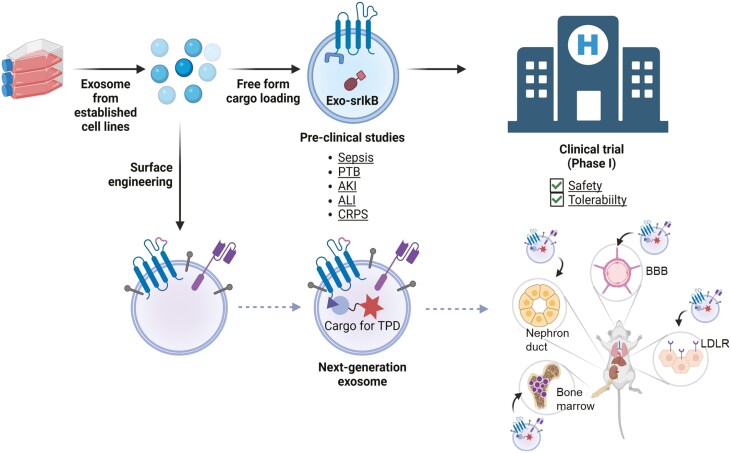
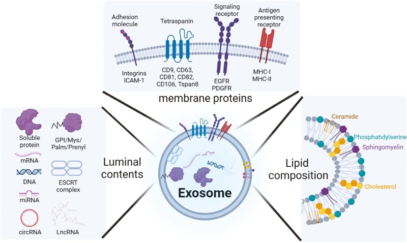
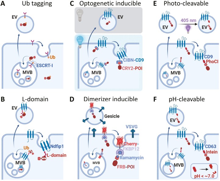
Extracellular vesicles, particularly exosomes, have emerged as promising drug delivery systems owing to their unique advantages, such as biocompatibility, immune tolerability, and target specificity. Various engineering strategies have been implemented to harness these innate qualities, with a focus on enhancing the pharmacokinetic and pharmacodynamic properties of exosomes via payload loading and surface engineering for active targeting. This concise review outlines the challenges in the development of exosomes as drug carriers and offers insights into strategies for their effective clinical translation. We also highlight preclinical studies that have successfully employed anti-inflammatory exosomes and suggest future directions for exosome therapeutics. These advancements underscore the potential for integrating exosome-based therapies into clinical practice, heralding promise for future medical interventions.

Keywords: drug delivery system; exosome purification; exosomes; extracellular vesicles; inflammation; protein therapeutics.


Conflict of interest statement

C.C. is the founder and shareholder; H.C. is an ex-employee and minor shareholder; and M.K., D.-J.J., and H. J. K., are employees of ILIAS Biologics, Inc. The authors declare no potential conflicts of interest.

Figures

Graphical Abstract
Graphical Abstract
Figure 1.
Figure 1.
Exosome constituents. Exosomes originate from endocytosis of lipid raft, resulting in a lipid composition similar to lipid raft. Local concentrations of cholesterol and sphingomyelin enable curvature of exosomal membrane, whereas phosphatidylserine located in the outer leaflet of the membrane creates negative charge of exosomes. Inverse cornical 3D conformation of tetraspanin also contributes to maintaining the curvature of exosomal membrane. In addition, adhesion molecules, as well as signaling receptors, confer exosomes binding preferences to certain types of cells. Exosomes contain a variety repertoire of proteins, nucleic acids, making them multi-functional molecules. Created with https://biorender.com/.
Figure 2.
Figure 2.
EV-loading strategies of free-cargo proteins. (A and B) involves the loading strategies mediated by ubiquitin-mediated interactions. (A) Ubiquitin tag, which is fused in the C-terminal tail of the cargo, is recognized by ESCRT complex for exosomal loading. (B) L-domain of an ILV marker protein interacts with a WW-domain in the cargo protein, facilitating mono ubiquitination of the cargo, followed by loading into intraluminal vesicles (ILVs). (C and D) utilizes light/chemical-induced protein–protein interaction (PPI). Photoactivated protein interactions occur between CRY-CIBN in the presence of light in (C) or FRB/FKBP in the presence of a rapamycin analog in (D). These reversible PPIs enable cargo loading into ILVs (one of PPI modules is connected to an ILV-enriched protein). Finally, (E and F) employ a protein-cleavable system. The cargo is covalently fused to the end of tetraspanin via photo- or pH-induced cleavage motif. In (E), the cargo is released from tetraspanin proteins by illumination of violet light to the purified EVs. In (F), the cargo is self-cleaved along with pH drop upon introduction into the ILVs. Created with https://biorender.com/.
Figure 3.
Figure 3.
Beagle scRNA sequencing results show the prominent effect of exosomes in neutrophils and monocytes among blood cells. (A and B) UMAP embedding of analyzed transcriptomes of 17 915 white blood cells colored by (A) the time point at which each cell was prepared after exosome infusion, (B) cell cluster groups along similarities in transcripts expression, and the cell types annotated with reference to human primary cell atlas. (A) 3251, 2688, 3568, 3105, 2867, and 2906 cells were prepared at 0 hour, 30 minutes, 1.5 hours, 3.5 hours, 6.5 hours, and 24 hours after exosome infusion. (B) Areas with black lines, monocyte-specific regions; an area with black dashed lines, region of monocytes mixed with other cell types. (C) A graph indicating cell types with the number of DEGs. (D) Expression level heatmap for statistically significant NF-κB signaling related factors of neutrophils. (E) Expression level heatmap for statistically significant NF-κB signaling related factors of monocytes. (F) Gene ontology (GO)-biological process (BP) terms of DEGs with significant changes in expression levels shown in (D). (G) Gene ontology (GO)-biological process (BP) terms of DEGs with significant changes in expression levels shown in (E).
Figure 4.
Figure 4.
Beagle scRNA sequencing results show the non-classical monocytes prominently affected by the exosome. (A) A bar plot indicating cell type proportion. Hematopoietic stem cells (HSCs). (B) Monocyte clusters are further grouped with classic or non-classic monocyte marker genes. (C) A bar plot indicating cell type proportion. (D) Expression level heatmap for statistically significant NF-κB signaling related factors of classical monocytes. (E) Expression level heatmap for statistically significant NF-κB signaling related factors of non-classical monocytes. (F) Venn diagram showing DEGs in monocytes, neutrophils, or NF-κB gene set. The numbers in parentheses are the number of DEGs from each population. (G) The proportions and expression patterns of 82 differentially expressed NF-κB genes in classical or non-classical monocytes are depicted as the size and color of the circles, respectively.

Similar articles

Cited by

References

    1. Li C, Wang J, Wang Y, et al.. Recent progress in drug delivery. Acta Pharm Sin B. 2019;9(6):1145-1162. 10.1016/j.apsb.2019.08.003 - DOI - PMC - PubMed
    1. Hua L, Zhang Q, Zhu X, et al.. Beyond proteolysis-targeting chimeric molecules: designing heterobifunctional molecules based on functional effectors. J Med Chem. 2022;65(12):8091-8112. 10.1021/acs.jmedchem.2c00316 - DOI - PubMed
    1. Liu YJ, Wang C.. A review of the regulatory mechanisms of extracellular vesicles-mediated intercellular communication. Cell Commun Signal. 2023;21(1):77. 10.1186/s12964-023-01103-6 - DOI - PMC - PubMed
    1. Sheller-Miller S, Choi K, Choi C, Menon R.. Cyclic-recombinase-reporter mouse model to determine exosome communication and function during pregnancy. Am J Obstet Gynecol. 2019;221(5): 502.e1-502.e12. 10.1016/j.ajog.2019.06.010 - DOI - PubMed
    1. Banks WA, Sharma P, Bullock KM, et al.. Transport of extracellular vesicles across the blood-brain barrier: brain pharmacokinetics and effects of inflammation. Int J Mol Sci. 2020;21(12):4407. 10.3390/ijms21124407 - DOI - PMC - PubMed