Review
. 2024 Jul;300(7):107444.
doi: 10.1016/j.jbc.2024.107444. Epub 2024 Jun 4.

To each its own: Mechanisms of cross-talk between GPI biosynthesis and cAMP-PKA signaling in Candida albicans versus Saccharomyces cerevisiae

Affiliations
Review

To each its own: Mechanisms of cross-talk between GPI biosynthesis and cAMP-PKA signaling in Candida albicans versus Saccharomyces cerevisiae

Sneha Sudha Komath. J Biol Chem. 2024 Jul.

Abstract

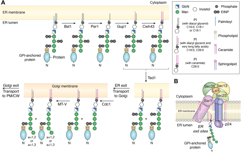
Candida albicans is an opportunistic fungal pathogen that can switch between yeast and hyphal morphologies depending on the environmental cues it receives. The switch to hyphal form is crucial for the establishment of invasive infections. The hyphal form is also characterized by the cell surface expression of hyphae-specific proteins, many of which are GPI-anchored and important determinants of its virulence. The coordination between hyphal morphogenesis and the expression of GPI-anchored proteins is made possible by an interesting cross-talk between GPI biosynthesis and the cAMP-PKA signaling cascade in the fungus; a parallel interaction is not found in its human host. On the other hand, in the nonpathogenic yeast, Saccharomyces cerevisiae, GPI biosynthesis is shut down when filamentation is activated and vice versa. This too is achieved by a cross-talk between GPI biosynthesis and cAMP-PKA signaling. How are diametrically opposite effects obtained from the cross-talk between two reasonably well-conserved pathways present ubiquitously across eukarya? This Review attempts to provide a model to explain these differences. In order to do so, it first provides an overview of the two pathways for the interested reader, highlighting the similarities and differences that are observed in C. albicans versus the well-studied S. cerevisiae model, before going on to explain how the different mechanisms of regulation are effected. While commonalities enable the development of generalized theories, it is hoped that a more nuanced approach, that takes into consideration species-specific differences, will enable organism-specific understanding of these processes and contribute to the development of targeted therapies.

Keywords: GPI-N-acetylglucosaminyltransferase; Ras; adenylyl cyclase; cAMP-PKA signaling; hyphae; pseudohyphae.


Conflict of interest statement

Conflict of interest The author declares that they have no conflicts of interest with the contents of this article.

Figures

Figure 1
Figure 1
The GPI biosynthetic pathway.A, formation of the complete GPI precursor and its attachment to proteins. The pathway shown here is from what is known in Saccharomyces cerevisiae. Not all steps have been elucidated in Candida albicans. However, homologs of most of the enzymes and their subunits are also present in C. albicans and the final GPI product is similar. GPI biosynthesis begins at the cytoplasmic face of the ER when GlcNAc from UDP-GlcNAc is transferred to PI by the action of a GlcNAc transferase (GPI-GnT). GlcNAc-PI is de-N-acetylated by the action of Gpi12 and then flipped into the ER lumen by the action of an unknown flippase. The first step on the lumenal face involves acylation (palmitoyl) by an acyltransferase, Gwt1. Subsequently four Man residues and three EtNP residues are transferred by the action of different mannosyltransferases (MT-I to MT-IV) and EtNP transferases (EtNPT-I to EtNPT-III), respectively, to generate the complete precursor (CP). The donor for the mannoses is Dol-P-Man and for EtNP is ethanolaminephosphate. The most likely sequential pathway alone is shown. Once CP is produced, it is amide linked to the C-terminal end of proteins by the action of a transamidase (GPIT). For this, GPIT cleaves the C-terminal GPI attachment signal sequence (SS) of proteins and attaches the EtNP on Man-3 of the CP to the newly generated carboxylate terminus. Inset: The nature of the GPI attachment signal sequence is conserved. The amino acids of the GPI attachment signal sequence are not conserved, but their nature is. These common features, mentioned in the figure, make it easy to identify them either manually or using bioinformatic tools. GPI attachment occurs at the ω residue. B, the known intra-subunit transcriptional cross talk within the C. albicans GPI-GnT and cross-talk with other biochemical pathways in the cell. Intersubunit transcriptional regulations within the GPI-GnT are unique to C. albicans and have not been reported in any other organism. Activations are represented by black arrows within the dotted circle representing the enzyme complex and the red flatheaded arrows represent repression within the GPI-GnT. Gpi15 activates the expression of both Gpi2 and Gpi19 while Eri1 represses both. Gpi15 and Eri1 mutually activate one another. Gpi2 and Gpi19 mutually inhibit the expression of one another and independently activate the expression of Gpi15. The roles of other GPI-GnT subunits in this schema are not yet elucidated. Cross-talk with other biochemical pathways is observed at this step. CaGpi19 and Erg11 (in sterol biosynthetic pathway) are mutually transcriptionally activated. Since Erg11 is the target of azoles, this affects the response of the cells to azole antifungals. CaGpi2 and CaEri1, in different ways, control filamentation via the Cyr1-cAMP-PKA pathway. CaGpi2 activates cAMP-PKA signaling for filamentation while CaEri1 represses it. The red flatheaded arrows represent inhibition and the blue arrows represent activation.
Figure 2
Figure 2
GPI remodeling and ER exit of GPI-anchored proteins using COPII vesicles.A, remodeling of the GPI anchor: The GPI anchor attached to proteins are remodeled in the ER lumen before they are transported out. Bst1 first deacylates the inositol. Per1 then removes the short chain fatty acid at sn2 position to produce a lyso-PI. Next, Gup1 attaches a very long chain fatty acid at this position. In a fraction of GPI-APs, the diacylglycerol may then be entirely replaced by a ceramide via the action of Cwh43. Finally, the EtNP on Man-2 is removed by Ted1. This step is crucial for recognition of the GPI-AP by the p24 proteins and their ER exit via COPII-coated vesicles. B, recruitment of p24 and COPII proteins for ER exit: In the lumen of the ER, GPI-APs containing remodeled very long chain lipid tails and lacking the EtNP on Man-2 accumulate at the ER exit sites. Here, they are recognized and bound by the p24 family of four membrane proteins. Once the cargo is bound, the cytoplasmic domains of the p24 proteins recruit Lst1 and Sec23, forming a pre-budding complex, which is also promoted by the association of Sar1, a membrane-bound GTPase, within this complex. The outer coat proteins, Sec13 and Sec31, can now assemble upon the pre-budding complex causing a resultant bending of the membrane and formation of nascent vesicles which are pinched off by the catalytic activity of Sar1.
Figure 3
Figure 3
The cAMP–PKA pathway that controls filamentation in Saccharomyces cerevisiae and Candida albicans. A schematic representation of the pathway in (A), S. cerevisiae and (B) C. albicans. PKA is a complex of the form R2C2 which activates filamentation. The R subunits are constituted by Bcy1 while the C subunits may be any two of Tpk1/Tpk2/Tpk3 in S. cerevisiae and Tpk1/Tpk2 in C. albicans. When cAMP is produced, it binds to Bcy1 and releases the active C subunits which phosphorylate downstream transcription factors and activate them. Depending on the C subunits released, the downstream targets vary. The production of cAMP is made possible by the adenylyl cyclase (Cyr1) which coverts ATP to cAMP. Any excess cAMP is degraded by phosphodiesterases (Pde2/Pde1). Pde2 (in bold) is the dominant player. Cyr1 is regulated by the binding of Ras2/Ras1 in S. cerevisiae and by Ras1 in C. albicans. It is also activated by Srv2/CAP1 and the binding of monomeric G-actin to the latter. In S. cerevisiae, heat shock proteins Hsc82 (or Hsp82) along with the co-chaperone, Sgt1, promote the interaction between Ras2 and Cyr1, whereas in C. albicans, Hsp90-Sgt1 inhibits Ras1–Cyr1 interaction. Ras proteins require to be in their active GTP-bound form for an effective interaction with Cyr1. For this, the GDP-bound inactive form of Ras must first be activated by the GEF, Cdc25 to the GTP-bound form. Ras cycles back to its inactive state by interacting with GAPs, Ira1/Ira2 in S. cerevisiae and Ira2 in C. albicans. This interaction can be blocked by a Gly19Val mutation in S. cerevisiae Ras2 and Gly13Val mutation in C. albicans Ras1, producing constitutively activated Ras proteins. PM localization of Ras proteins is dependent on their farnesylation in the cytoplasm and palmitoylation at the ER. Gpr1-Gpa2 is a G-protein–coupled receptor-G-protein pair required to transport glucose (S. cerevisiae) or lactose/Met (C. albicans). Phosphorylated Gpa2 can activate Cyr1. The conversion of glucose to fructose 1,6-bis-phosphate (Fru1,6bisP) activates Cdc25 and turns on Ras-dependent Cyr1-cAMP-PKA signaling in S. cerevisiae. It is hypothesized that a similar mechanism operates in C. albicans. CO2-mediated activation of Cyr1 is exclusive to C. albicans and occurs independent of Ras1–Cyr1 interaction. Double headed arrows represent protein–protein interactions, the red flatheaded arrows represent inhibition, and the blue arrows represent activation.
Figure 4
Figure 4
Domain organization of Cyr1 in Saccharomyces cerevisiae and Candida albicans. A schematic representation of the domains of Cyr1 from (A) S. cerevisiae and (B) C. albicans. Cyr1 is the adenylyl cyclase that converts ATP to cAMP and thereby activates PKA. This enzymatic activity primarily requires the cyclase domain of Cyr1. The other domains act as sensors of different proteins/signals to regulate cAMP production by the cyclase domain. The Gα-binding domain (GαBD) binds to the α subunit (Gpa2) of a G-protein; Ras association (RA) domain interacts with Ras proteins; the leucine-rich repeat (LRR) domain interacts with Hsp90 proteins via their co-chaperone, Sgt1; and the cyclase associated protein 1 (CAP1) binding domain (CBD) interacts with Srv2/CAP1 protein. The role of the protein phosphatase 2C (PP2C) domain is not known. In C. albicans, the LRR domain is activated by bacterial peptidoglycans and muramyl dipeptides (shown by bluearrow). Its cyclase domain can be activated independently by CO2 (shown by bluearrow) and repressed by farnesol (represented by red flatheaded arrow). Hyperactive mutants of C. albicans Cyr1 (Cyr1E1541K) can simultaneously bind Ras1 and CAP1. Double headed arrows represent protein–protein interactions.
Figure 5
Figure 5
Ras proteins of Saccharomyces cerevisiae and Candida albicans.A, post-translational modifications of Ras proteins and their subcellular localization. Ras proteins are produced in the cytoplasm. The N-terminal Met is removed almost immediately by methionine aminopeptidase and a farnesyl chain is added to the Cys residue of the C-terminal CAAX (C: Cys, A: aliphatic amino acids, X: the C-terminal amino acid) domain by a farnesyltransferase. The AAX motif is then proteolytically cleaved by an endopeptidase, Rce1, and the new C-terminal -COOH generated is converted to an ester by the action of an isoprenylcysteine methyltransferase (ICMT). Finally, a palmitoyltransferase enzyme complex adds a palmitoyl chain to an adjacent Cys and the protein is transported to the PM. B, domain organization of mature ScRas2 and CaRas1. Mature Ras proteins contain five well-conserved G boxes at their N-terminus that are required for GTP binding. The G1 and G2 motifs bind Mg+2-bound β and γ Pi, respectively. The crucial Gln required for GTPase activity is located in the G3 motif. G4 and G5 motifs bind guanine and the ribose sugar. Nucleotide exchange after GEF binding induces large conformational changes in switch I and switch II regions. ScRas2/CaRas1 also have hypervariable regions (HVR) whose sequences are poorly conserved. The C-terminal CAAX domain and the residues on which farnesylation and palmitoylation occur are specifically shown.
Figure 6
Figure 6
Cross-talk of GPI-GnT with Cyr1-cAMP-PKA pathway in Saccharomyces cerevisiae and Candida albicans. Shown in the figure are probable models of the cross-talk in (A) S. cerevisiae and (B) C. albicans. Ras proteins interact with the GPI-GnT at the ER during their transit to the PM. How this transition occurs is shown in Figure 5. This requires active Ras and, at least in S. cerevisiae, the effector loop. The interaction leads to inhibition of the GPI-GnT in S. cerevisiae and its activation in C. albicans. In C. albicans, only the Gpi2 subunit physically interacts with Ras. Perhaps the same subunit interacts with Ras in S. cerevisiae, but no clear-cut evidence exists for this. The GPI-GnT in S. cerevisiae inhibits the Cyr1-cAMP-PKA signaling. One possible model would be to assume that when bound to the GPI-GnT in the ER, the molecules of Ras available at the PM to activate cAMP production drop. This leads to the inhibition of filamentation. Downregulation of any of the GPI-GnT subunits releases Ras and activates filamentation. In C. albicans, overexpression of Gpi2 inhibits Hsp90, whose downregulation enables better interaction of Ras with Cyr1 and activates filamentation. If such an effect were to operate in S. cerevisiae, it would lead to inhibition of filamentation, since Hsc82 promotes the interaction of Ras with Cyr1 in this organism. But evidence in support of such a model is lacking. Filamentation in C. albicans is also activated by the direct binding of CO2 to the cyclase domain of Cyr1, independent of Ras. The Eri1 subunit of the GPI-GnT inhibits this process. Double headed arrows represent protein–protein interactions, red flatheaded arrows represent inhibition, and blue arrows represents activation.

Similar articles

References

    1. Komath S.S., Fujita M., Hart G.W., Ferguson M.A.J., Kinoshita T. In: Essentials of Glycobiology. 4th Ed. Varki A., Cummings R.D., Esko J.D., Stanley P., Hart G.W., Aebi M., et al., editors. Cold Spring Harbor Laboratory Press; Cold Spring Harbor, NY: 2022. Glycosylphosphatidylinositol anchors.
    1. Kinoshita T. Biosynthesis and biology of mammalian GPI-anchored proteins. Open Biol. 2020;10 - PMC - PubMed
    1. Shukla N., Tang W.K., Tolia N.H. Structural analysis of Plasmodium falciparum ookinete surface antigen Pfs28 relevant for malaria vaccine design. Sci. Rep. 2022;12 - PMC - PubMed
    1. Karim M., Singh G., Thakur S., Rana A., Rub A., Akhter Y. Evaluating complete surface-associated and secretory proteome of Leishmania donovani for discovering novel vaccines and diagnostic targets. Arch. Microbiol. 2022;204:604. - PubMed
    1. Pappas P.G., Vazquez J.A., Oren I., Rahav G., Aoun M., Bulpa P., et al. Clinical safety and efficacy of novel antifungal, fosmanogepix, for the treatment of candidaemia: results from a phase 2 trial. J. Antimicrob. Chemother. 2023;78:2471–2480. - PMC - PubMed

Publication types

MeSH terms

Substances

LinkOut - more resources