

Digital ray: enhancing cataractous fundus images using style transfer generative adversarial networks to improve retinopathy detection
- PMID: 38839251
- PMCID: PMC11503040
- DOI: 10.1136/bjo-2024-325403
Digital ray: enhancing cataractous fundus images using style transfer generative adversarial networks to improve retinopathy detection
Abstract
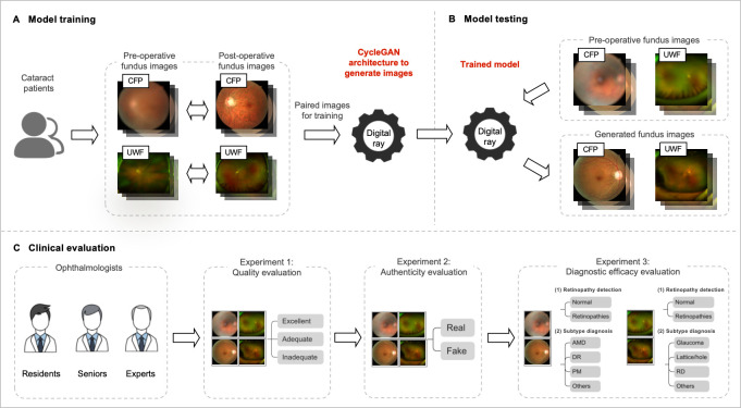
Background/aims: The aim of this study was to develop and evaluate digital ray, based on preoperative and postoperative image pairs using style transfer generative adversarial networks (GANs), to enhance cataractous fundus images for improved retinopathy detection.
Methods: For eligible cataract patients, preoperative and postoperative colour fundus photographs (CFP) and ultra-wide field (UWF) images were captured. Then, both the original CycleGAN and a modified CycleGAN (C2ycleGAN) framework were adopted for image generation and quantitatively compared using Frechet Inception Distance (FID) and Kernel Inception Distance (KID). Additionally, CFP and UWF images from another cataract cohort were used to test model performances. Different panels of ophthalmologists evaluated the quality, authenticity and diagnostic efficacy of the generated images.
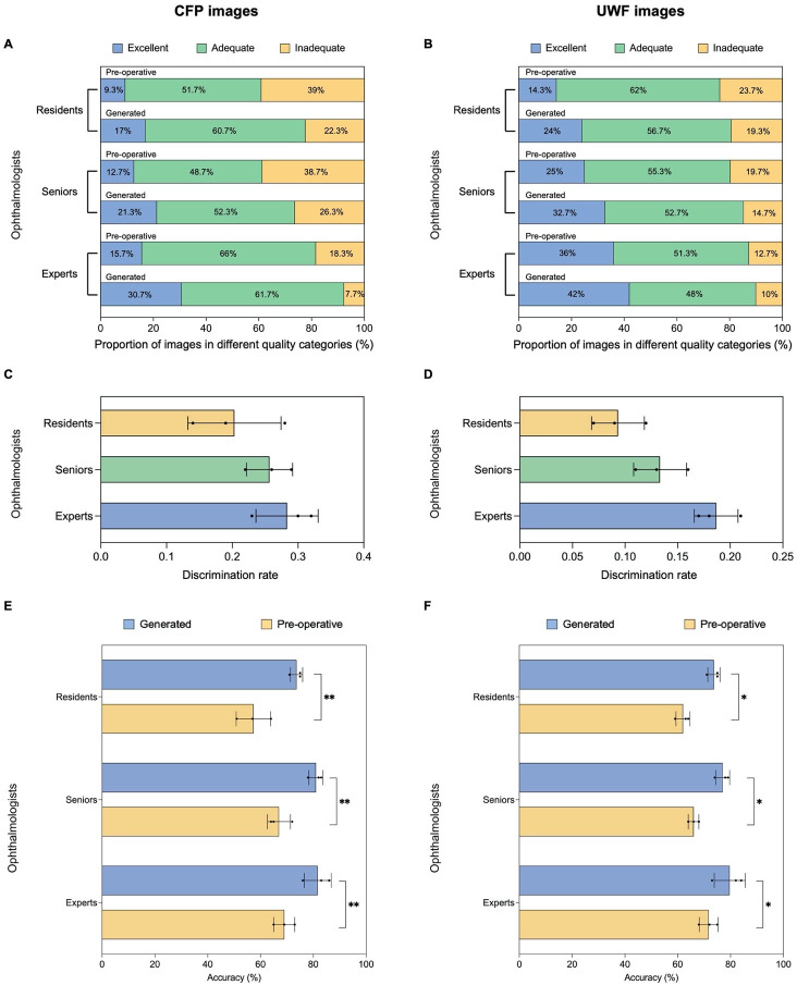
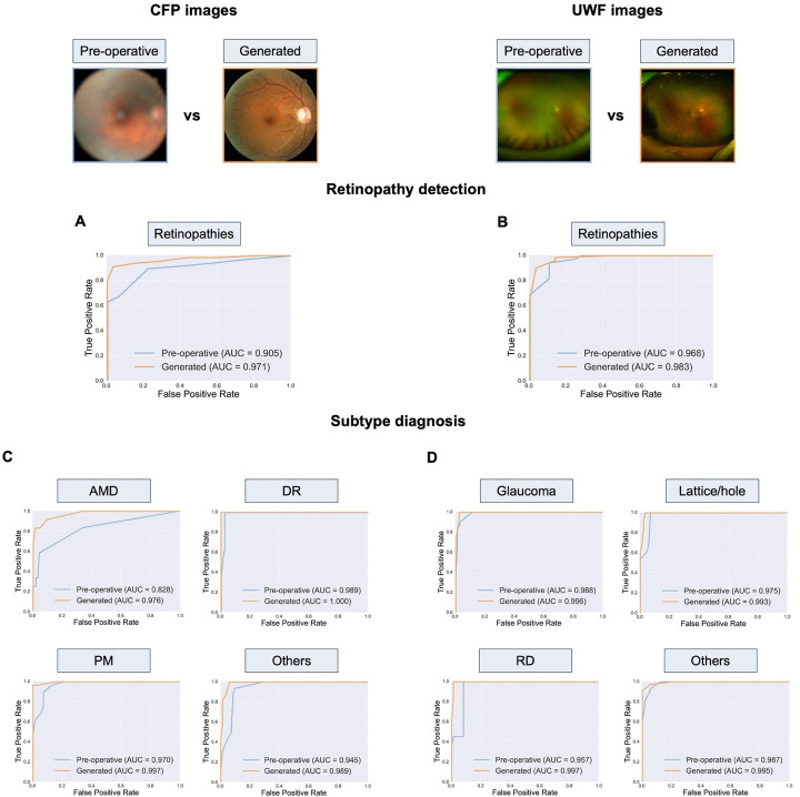
Results: A total of 959 CFP and 1009 UWF image pairs were included in model development. FID and KID indicated that images generated by C2ycleGAN presented significantly improved quality. Based on ophthalmologists' average ratings, the percentages of inadequate-quality images decreased from 32% to 18.8% for CFP, and from 18.7% to 14.7% for UWF. Only 24.8% and 13.8% of generated CFP and UWF images could be recognised as synthetic. The accuracy of retinopathy detection significantly increased from 78% to 91% for CFP and from 91% to 93% for UWF. For retinopathy subtype diagnosis, the accuracies also increased from 87%-94% to 91%-100% for CFP and from 87%-95% to 93%-97% for UWF.
Conclusion: Digital ray could generate realistic postoperative CFP and UWF images with enhanced quality and accuracy for overall detection and subtype diagnosis of retinopathies, especially for CFP.\ TRIAL REGISTRATION NUMBER: This study was registered with ClinicalTrials.gov (NCT05491798).
Keywords: Imaging; Lens and zonules; Retina.
© Author(s) (or their employer(s)) 2024. Re-use permitted under CC BY-NC. No commercial re-use. See rights and permissions. Published by BMJ.
Conflict of interest statement
Competing interests: None declared.
Figures
Similar articles
-
Deep learning-based classification of retinal vascular diseases using ultra-widefield colour fundus photographs.BMJ Open Ophthalmol. 2022 Feb 4;7(1):e000924. doi: 10.1136/bmjophth-2021-000924. eCollection 2022. BMJ Open Ophthalmol. 2022. PMID: 35141420 Free PMC article.
-
ETDRS grading with CLARUS ultra-widefield images shows agreement with 7-fields colour fundus photography.BMC Ophthalmol. 2024 Sep 3;24(1):387. doi: 10.1186/s12886-024-03537-z. BMC Ophthalmol. 2024. PMID: 39227901 Free PMC article.
-
Comparison of widefield swept-source optical coherence tomography angiography with ultra-widefield colour fundus photography and fluorescein angiography for detection of lesions in diabetic retinopathy.Br J Ophthalmol. 2021 Apr;105(4):577-581. doi: 10.1136/bjophthalmol-2020-316245. Epub 2020 Jun 26. Br J Ophthalmol. 2021. PMID: 32591347 Free PMC article.
-
Update on wide- and ultra-widefield retinal imaging.Indian J Ophthalmol. 2015 Jul;63(7):575-81. doi: 10.4103/0301-4738.167122. Indian J Ophthalmol. 2015. PMID: 26458474 Free PMC article. Review.
-
Ultra-Widefield Imaging for Evaluation of the Myopic Eye.Semin Ophthalmol. 2021 May 19;36(4):185-190. doi: 10.1080/08820538.2021.1887904. Epub 2021 Feb 23. Semin Ophthalmol. 2021. PMID: 33620294 Review.
References
-
- A human-centered evaluation of a deep learning system deployed in clinics for the detection of diabetic retinopathy. Proceedings of the 2020 CHI Conference on Human Factors in Computing Systems; 2020.
Publication types
MeSH terms
Associated data
LinkOut - more resources
Full Text Sources
Medical
Miscellaneous