Review
. 2024 Jun 5;44(23):e2104232024.
doi: 10.1523/JNEUROSCI.2104-23.2024.

Celebrating the Birthday of AMPA Receptor Nanodomains: Illuminating the Nanoscale Organization of Excitatory Synapses with 10 Nanocandles

Affiliations
Review

Celebrating the Birthday of AMPA Receptor Nanodomains: Illuminating the Nanoscale Organization of Excitatory Synapses with 10 Nanocandles

Yuko Fukata et al. J Neurosci. .

Abstract

A decade ago, in 2013, and over the course of 4 summer months, three separate observations were reported that each shed light independently on a new molecular organization that fundamentally reshaped our perception of excitatory synaptic transmission (Fukata et al., 2013; MacGillavry et al., 2013; Nair et al., 2013). This discovery unveiled an intricate arrangement of AMPA-type glutamate receptors and their principal scaffolding protein PSD-95, at synapses. This breakthrough was made possible, thanks to advanced super-resolution imaging techniques. It fundamentally changed our understanding of excitatory synaptic architecture and paved the way for a brand-new area of research. In this Progressions article, the primary investigators of the nanoscale organization of synapses have come together to chronicle the tale of their discovery. We recount the initial inquiry that prompted our research, the preceding studies that inspired our work, the technical obstacles that were encountered, and the breakthroughs that were made in the subsequent decade in the realm of nanoscale synaptic transmission. We review the new discoveries made possible by the democratization of super-resolution imaging techniques in the field of excitatory synaptic physiology and architecture, first by the extension to other glutamate receptors and to presynaptic proteins and then by the notion of trans-synaptic organization. After describing the organizational modifications occurring in various pathologies, we discuss briefly the latest technical developments made possible by super-resolution imaging and emerging concepts in synaptic physiology.


Conflict of interest statement

The authors declare no competing financial interest.

Figures
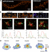
Figure 1.
Figure 1.
Discovery of AMPAR nanodomains by using various super-resolution techniques. A, Live-cell sptPALM imaging with GluA2-mEos overexpression (left) and fixed STED imaging on endogenous GluA2 (right) example. B, Zoom on spines where GluA2 nanodomains are revealed with sptPALM, STED, and uPAINT techniques. C, Example of 2C STORM with endogenous PSD-95 (red) and AMPAR (green) with a line scan to quantify each signal intensity. D, Examples of PSDs resolved with PALM for mEos2-tagged shrPSD-95, GKAP, shrShank3, and Homer1c, showing that the major PSD scaffold molecules are organized in subsynaptic nanodomains. Scale bar: 200 nm. Figure panels (A–C) are reproduced from Nair et al. (2013) and (D) from MacGillavry et al. (2013).
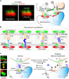
Figure 2.
Figure 2.
Discovery of postsynaptic nanodomains based on the palmitoylation study. A,B, Live-cell STED imaging (green), but not conventional confocal microscopy (red pseudocolor), of neurons expressing PF11-GFP, an intrabody against palmPSD-95, apparently detects postsynaptic nanodomains (1–4 nanodomains/spine). C, 2C-STED (upper panel) analysis of fixed neurons shows that postsynaptic PSD-95 nanodomains (red pseudocolor, arrows) are covered by a single presynaptic bouton (vGlut1 green pseudocolor). The same fields are sequentially imaged in the confocal mode (lower). D, 2C-STED analysis of live neurons reveals that individual PSD-95 nanodomains (red pseudocolor, arrows) are associated with smaller clusters of surface-expressed AMPAR subunit, GluA1 (green pseudocolor). Asterisks indicate extrasynaptic AMPARs. E, Hippocampal neurons treated with 90 mM KCl for 5 min were stained by PSD-95 and Bassoon antibodies and visualized by STED microscopy. Arrows indicate PSD-95 signals observed away from the synaptic sites upon high K + treatment. F, Model for PSD-95 nanodomain formation in excitatory postsynapses by local palmitoylation cycles. palmPSD-95 is partitioned into discrete nanodomains (two red ovals) to constitute a PSD. Spine membrane-inserted zDHHC-palmitoylating enzymes and ABHD17-depalmitoylating enzymes locally generate PSD-95 palmitoylation cycles to prevent lateral diffusion of membrane-bound PSD-95 and maintain the individual nanodomains. Figure panels (A–E) are reproduced from Fukata et al. (2013).
Figure 3.
Figure 3.
A trans-synaptic nanocolumn and nanopathology. A, Trans-synaptic nanocolumns composed of pre- and postsynaptic nanodomains. 2C-STED imaging of Bassoon (green pseudocolor) and palmPSD-95 (red pseudocolor) suggests two nanocolumns in a presented synapse. VDCC, voltage-dependent calcium channel. This figure is reproduced from Fukata and Fukata (2010). B, Proposed nanocolumn constituents including Nrxn−Nlgn, Nrxn−LRRTM, Nrxn−Cbln−GluD, and LGI1−ADAM22/23. +S4 and −S4 indicate Nrxns with or without the splice site 4 insert, respectively. C, Nanocolumns mediated by LGI1–ADAM22–PSD-95. Left panels, 2C-STED imaging unveils that extracellularly labeled ADAM22 (red) and LGI1 (green) coincided in nanoclusters, which are closely flanked by presynaptic Bassoon and postsynaptic PSD-95 (green) in the hippocampal CA1 region (modified from Fukata et al., 2021). Right diagram, LGI1–ADAM22–PSD-95 interaction plays a critical role in finely regulated AMPAR-mediated synaptic transmission and physiological brain excitability. If the interaction in LGI1–ADAM22–PSD-95 is disrupted for some reasons, such as congenital mutations (indicated by “X”) or acquired autoantibodies (red, “Y shape“), epilepsy and/or memory impairment occur(s) in our brain (right) (modified from Fukata et al., 2021).
Figure 4.
Figure 4.
Nanoscale map of APP in functional zones of the synapse (Kedia et al., 2020, A, Nanoscale localization of the C-terminal domain of APP (magenta) with the PSD (green). The overlay is denoted in black. The top row shows a confocal image, the middle row displays super-resolution images, and the bottom row illustrates diffraction-limited PSD in black, super-resolved PSD in green and nanodomains of APP in magenta. B, Nanoscale localization of the C-terminal domain of APP (magenta) with the endocytic zone (green). The overlay is denoted in black. The top row shows a confocal image, the middle row displays super-resolution images, and the bottom row illustrates the diffraction-limited endocytic zone in black, super-resolved endocytic zone in green and nanodomains of APP in magenta. Scale bar: 600 nm. C, Data-driven nanoscale map of the amyloidogenic machinery in a dendritic spine. APP (substrate) and β- and γ-secretases (canonical secretases) are coded as red, green, and purple, respectively. The central zone represents PSD, the peripheral annular zone represents the endocytic zone, and the rest of the extrasynaptic compartment is depicted in yellow.
Figure 5.
Figure 5.
Synaptic scaffolding proteins form nanoscale condensates at excitatory synapses (Narayanan et al., 2019 ; Rajeev et al., 2022). A, In vitro condensation of the C-terminal supramolecular region of SAP97 isoforms into micron-sized condensates in the presence of a crowding agent. B, Nanoscale condensates of SAP97 observed in the dendritic shaft and spines of pyramidal neurons. Left top, epifluorescence; middle, super-resolved; bottom, overlay. The inset indicates a zoomed-in version of selected regions of interest. Scale bars, 5 µm; insets, 1.5 µm. C, Mutually exclusive formation of nano-organization of PSD-95 (magenta) and SAP97 (green) at excitatory synapses (right panel) and PSD (PSD-95, black region) and nanocondensates of SAP97 (pseudo color) in the left panel. D, A plot showing the probability distribution function of single molecules detected inside the nanoclusters. E, The curve fit of the inverse of the probability distribution of molecules, following the function an^2/3 − bn + c, to obtain the parameters a, b, and c, which define the nucleation barrier (ΔGc) and critical cluster radius (Rc). F, A schematic representation of the free energy function, illustrating a first-order phase transition. The broad arrows represent the favored direction of aggregate size concerning spontaneous growth or diffusion of aggregates. The solid line represents the extent of experimental values.

Similar articles

Cited by

References

    1. Baddeley D, Crossman D, Rossberger S, Cheyne JE, Montgomery JM, Jayasinghe ID, Cremer C, Cannell MB, Soeller C (2011) 4D super-resolution microscopy with conventional fluorophores and single wavelength excitation in optically thick cells and tissues. PLoS One 6:e20645. 10.1371/journal.pone.0020645 - DOI - PMC - PubMed
    1. Bartol TM, Keller DX, Kinney JP, Bajaj CL, Harris KM, Sejnowski TJ, Kennedy MB (2015) Computational reconstitution of spine calcium transients from individual proteins. Front Synaptic Neurosci 7:17. 10.3389/fnsyn.2015.00017 - DOI - PMC - PubMed
    1. Bats C, Groc L, Choquet D (2007) The interaction between stargazin and PSD-95 regulates AMPA receptor surface trafficking. Neuron 53:719–734. 10.1016/j.neuron.2007.01.030 - DOI - PubMed
    1. Baude A, Nusser Z, Roberts JD, Mulvihill E, McIlhinney RA, Somogyi P (1993) The metabotropic glutamate receptor (mGluR1 alpha) is concentrated at perisynaptic membrane of neuronal subpopulations as detected by immunogold reaction. Neuron 11:771–787. 10.1016/0896-6273(93)90086-7 - DOI - PubMed
    1. Bernard V, Somogyi P, Bolam JP (1997) Cellular, subcellular, and subsynaptic distribution of AMPA-type glutamate receptor subunits in the neostriatum of the rat. J Neurosci 17:819–833. 10.1523/JNEUROSCI.17-02-00819.1997 - DOI - PMC - PubMed

Substances

LinkOut - more resources