

Topical Solution for Retinal Delivery: Bevacizumab and Ranibizumab Eye Drops in Anti-Aggregation Formula (AAF) in Rabbits
- PMID: 38839719
- PMCID: PMC11196329
- DOI: 10.1007/s11095-024-03721-2
Topical Solution for Retinal Delivery: Bevacizumab and Ranibizumab Eye Drops in Anti-Aggregation Formula (AAF) in Rabbits
Abstract
Purpose: Wet age-related macular degeneration (AMD) is a blinding retinal disease. Monthly intravitreal anti-VEGF antibody injections of bevacizumab (off-label) and ranibizumab (FDA approved) are the standard of care. Antibody aggregation may interfere with ocular absorption/distribution. This study assessed topical delivery of dilute antibodies to the posterior segment of rabbit eyes using a novel anti-aggregation formula (AAF).
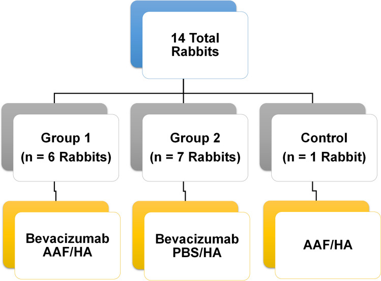
Methods: Bevacizumab, or biosimilar ranibizumab was diluted to 5 mg/ml in AAF. All rabbits were dosed twice daily. Substudy 1 rabbits (bevacizumab, 100 µl eye drops): Group 1 (bevacizumab/AAF, n = 6); Group 2 (bevacizumab/PBS, n = 7) and Vehicle control (AAF, n = 1). Substudy 2 rabbits (ranibizumab biosimilar/AAF, 50 µl eye drops): (ranibizumab biosimilar/AAF, n = 8). At 14.5 days, serum was drawn from rabbits. Aqueous, vitreous and retina samples were recovered from eyes and placed into AAF aliquots. Tissue analyzed using AAF as diluent.
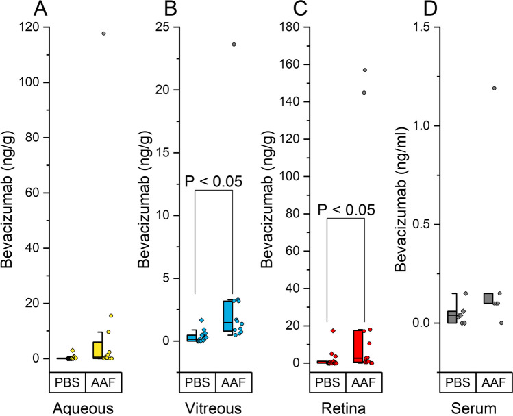
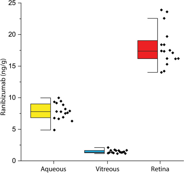
Results: Bevacizumab in AAF permeated/accumulated in rabbit aqueous, vitreous and retina 10 times more, than when diluted in PBS. AAF/0.1% hyaluronic acid eye drops, dosed twice daily, provided mean tissue concentrations (ng/g) in retina (29.50), aqueous (12.34), vitreous (3.46), and serum (0.28 ng/ml). Additionally, the highest concentration (ng/g) of ranibizumab biosimilar was present in the retina (18.0), followed by aqueous (7.82) and vitreous (1.47). Serum concentration was negligible (< 0.04 ng/ml). No irritation was observed throughout the studies.
Conclusions: Bevacizumab and ranibizumab, in an AAF diluent eye drop, can be delivered to the retina, by the twice daily dosing of a low concentration mAb formulation. This may prove to be an adjunct to intravitreal injections.
Keywords: anti-VEGF; antibody; macular degeneration; neovascularization; retina.
© 2024. The Author(s).
Conflict of interest statement
The authors Steven A. Giannos and Edward R. Kraft own significant interest in the IP of the technology described. The authors Jonathan D. Luisi, Mary E. Schmitz-Brown, Valentina Reffatto, Kevin H. Merkley and Praveena K. Gupta have no competing interests.
Figures
References
MeSH terms
Substances
LinkOut - more resources
Full Text Sources
