. 2024 Jun 5;15(1):4781.
doi: 10.1038/s41467-024-49025-2.

A candidate sex determination locus in amphibians which evolved by structural variation between X- and Y-chromosomes

Affiliations

A candidate sex determination locus in amphibians which evolved by structural variation between X- and Y-chromosomes

Heiner Kuhl et al. Nat Commun. .

Abstract

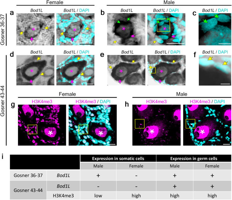
Most vertebrates develop distinct females and males, where sex is determined by repeatedly evolved environmental or genetic triggers. Undifferentiated sex chromosomes and large genomes have caused major knowledge gaps in amphibians. Only a single master sex-determining gene, the dmrt1-paralogue (dm-w) of female-heterogametic clawed frogs (Xenopus; ZW♀/ZZ♂), is known across >8740 species of amphibians. In this study, by combining chromosome-scale female and male genomes of a non-model amphibian, the European green toad, Bufo(tes) viridis, with ddRAD- and whole genome pool-sequencing, we reveal a candidate master locus, governing a male-heterogametic system (XX♀/XY♂). Targeted sequencing across multiple taxa uncovered structural X/Y-variation in the 5'-regulatory region of the gene bod1l, where a Y-specific non-coding RNA (ncRNA-Y), only expressed in males, suggests that this locus initiates sex-specific differentiation. Developmental transcriptomes and RNA in-situ hybridization show timely and spatially relevant sex-specific ncRNA-Y and bod1l-gene expression in primordial gonads. This coincided with differential H3K4me-methylation in pre-granulosa/pre-Sertoli cells, pointing to a specific mechanism of amphibian sex determination.


Conflict of interest statement

The authors declare no competing interests.

Figures

Fig. 1
Fig. 1. The chromosome-scale reference-genome of Bufo(tes) viridis, its karyotype and comparison with the reference genome of Bufo bufo.
a Breeding pair of B. viridis in amplexus (© M. Stöck). b Giemsa-stained karyotype of B. viridis (© M. Stöck). c B. viridis-assembly (y-axis) compared to B. bufo (x-axis) assembly by the Vertebrate Genome Project (VGP, GenBank accession number GCA_905171765.1) showing high synteny of both bufonid genomes across all 11 chromosomes.
Fig. 2
Fig. 2. The sex-diagnostic marker of Bufo(tes) viridis and its diploid relatives among Palearctic green toads.
a top: Mapping the pool-seq short reads to the B. viridis reference showed genome-wide a single region on scaffold 1, where the number of heterozygous SNPs (y-axis) (N = 103) in males surpassed about nine times the strongest signal of female heterozygous SNPs (N = 12), provides strong evidence for male heterogamety (XX/XY; for a putative ZZ/ZW system: Supplementary Fig. 3); a middle: male heterozygous SNPs on scaffold 1 of the male genome assembly identifies the same region (note a slight shift of the peak showing the same locus due to different reference genomes); a bottom: male-specific pool-seq ('pool-sex') coverage on the male genome (for the entire female and male genomes: Supplementary Data 6); y-axis for heterozygous SNPs are counts of SNPs (above and middle subfigure) or Y-specifically pool-sex covered base pairs (lower subfigure) in a cluster, where the maximal distance of variants is 9000 bp. b Representative sex-specific PCR-products of primers BvXY1 in females (♀♀, left) and males (♂♂, right) of six (i-vi) nominal diploid green toad taxa, electrophoresis result using 4 μL of PCR-products on a 4% high-resolution Sieve-agarose gel run for 60 min on 70 V; examined numbers of females (♀) and males (♂) for Palearctic green toad taxa were: (i) siculus (4♀, 6♂), (ii) shaartusiensis (highest band shown in males is weak; 4♀, 5♂), (iii) viridis and (iv) variabilis (49♀, 44♂), (v) balearicus (6♀, 4♂), (vi) turanensis (6♀, 4♂). c Range map of Palearctic green toad taxa (based on mitochondrial DNA and ploidy inferred from karyotypes, flow cytometry and nuclear microsatellite genotypes), numbers (i-vi) and color-coding as in (b) and (e), diploid (2n) taxa are indicated by circles, triploids (3n) by triangles and tetraploids (4n) by squares (map ca. 11° W to 97° E, 25° N to 62° N). d FISH-TSA on a metaphase spread from a B. viridis male with red bod1l-signals on chromosomes 1 on their short (p) arm (arrows), chromosomes were counterstained with DAPI (blue-green), scale bar in lower left corner represents 10 µm (ca. 40 metaphases were inspected). e Dated mitochondrial phylogeny of Palearctic green toads (adapted from ref. , with the phylogenetic positions of the diploid taxa, in which BvXY1 is sex-diagnostic, indicated by color-coded arrows as in (b, c); the three ploidy levels are provided to the right of the leaves, note that the sex marker is seemingly conserved only in the diploid green toads; all polyploids are of hybrid origin.
Fig. 3
Fig. 3. Additive sex-specific XX- or XY-expression of bod1l and expression of the ncRNA-Y in females and males in seven developmental stages, with numbers (N) of females and males (N) per stage shown in bars, genetically sexed using PCR-marker BvXY1.
Gosner stages 23–24, 10 days after fertilization, Gosner stages 27–29: 15 days after fertilization, Gosner stages 30–33: 18 days after fertilization, Gosner stages 34–38: 34 days after fertilization, Gosner stages 43–44: beginning of metamorphosis, Gosner stages 45–46, including toads up to six weeks after metamorphosis, the latter with anatomically visible differentiation of testes and Bidder’s organs in males, and ovaries in females, subadult male and adult female. Numbers of available tadpole transcriptomes varied by stage; TPM: Transcript reads Per Million RNAseq reads; note that TPM for the ncRNA-Y were multiplied 20x. See Supplementary Fig. 8 for comparison of additive sex-specific expression of bod1l and ncRNA-Y and haplotype-specific bod1l-expression in males. Bars present mean values; error bars show the standard deviation of the respective data points.
Fig. 4
Fig. 4. Structure of the X- and Y-haplotype of the candidate sex locus of Bufo(tes) viridis with focus on the 5′-end of bod1l and the Y-specific ncRNA.
a X-chromosomal (above, pink) and Y-chromosomal haplotype (below, blue) with color-coded homologous or X- and Y-specific INDELs, and the Y-specific long (ca. 5.8 kb) non-coding RNA (ncRNA-Y), flanking a putative enhancer at the 5′-end of the gene, ca. 80 kilobases before its promoter. b Graph of the signal of the AI-based software i-enhancer, reaching from 0 to 2, predicting strong enhancer properties of the Y-specific 5′-end of bod1l, including a G-quadruplex-rich region on the non-template strand, overlapping with the ncRNA-Y. c Y- and thus male-specific expression of the ncRNA-Y (204 bp PCR-product from cDNA) in larval green toads, from 10 days after fertilization (Gosner stages 23–24) until early metamorphosis (Gosner 46); in females, only non-specific products occurred, since X-copies of bod1l do not contain this ncRNA (for details and positive controls of females: Supplementary Fig. 9). d Unrooted Maximum-Likelihood-based phylogenetic tree, showing that the ncRNA-Y evolved in a common ancestor of diploid Palearctic green toads sharing this XY-system, with siculus and viridis spanning the most divergent lineages; two PCR-products (2.5 and 5.8 kb), directly amplified by independent PCRs from six related diploid green toad taxa, clustered by Y-gametologue and thus species (* symbolized 100% bootstrap support; only support values > 50% are shown).
Fig. 5
Fig. 5. Bod1l-expression and H3K4me3-methylation in primordial gonads of B. viridis, i.e., in the female early ovary, and in male Bidder’s organ, contributing to the testes anlage (Gosner stages 36–37) as well as in early metamorphosis (Gosner stages 43–44).
Bod1l expression shown by RNA in-situ hybridization (ISH) on 20 µm transverse cryosections of female (a, d) and male (b, c, e, f, h) gonads at developmental stages Gosner 36–37 (ac) and 43–44 (df), the latter presenting early metamorphic stages. DAPI was used to stain nuclei. Magenta asterisks indicate nuclei of germ cells; magenta arrows: cytoplasm of germ cells; yellow arrows: nuclei of somatic cells not stained by bod1l RNA ISH; green arrows: nuclei of somatic cells stained by bod1l RNA ISH. Yellow boxes in b and e indicate zoomed images shown in c and f, respectively. H3K4me3 and DAPI staining on 20 µm transverse cryosections of female (g) and male (h) green toad Bidder’s organ and gonads, Gosner stage 43–44. White asterisks indicate H3K4me3-positive germ cell nuclei; yellow boxes indicate somatic cells. Scale bars (a, b, d, e, g, h): 20 µm; (c, f) 5 µm. i Summary of bod1l-expression and H3K4me3-methylation in somatic cells and germ cells in Gosner stages 36–37 and 43–44 male and female gonads. For each Gosner stage, five sections from one gonad sample were analyzed in two independent experiments.

Similar articles

Cited by

References

    1. Herpin A, Schartl M. Plasticity of gene-regulatory networks controlling sex determination: of masters, slaves, usual suspects, newcomers, and usurpators. EMBO Rep. 2015;16:1260–1274. doi: 10.15252/embr.201540667. - DOI - PMC - PubMed
    1. Capel B. Vertebrate sex determination: evolutionary plasticity of a fundamental switch. Nat. Rev. Genet. 2017;18:675–689. doi: 10.1038/nrg.2017.60. - DOI - PubMed
    1. Bachtrog D, et al. Sex determination: why so many ways of doing it? PLoS Biol. 2014;12:e1001899. doi: 10.1371/journal.pbio.1001899. - DOI - PMC - PubMed
    1. Rovatsos M, et al. Conservation of sex chromosomes in lacertid lizards. Mol. Ecol. 2016;25:3120–3126. doi: 10.1111/mec.13635. - DOI - PubMed
    1. Beukeboom, L. W. & Perrin, N. The evolution of sex determination (Oxford University Press, 2014).