. 2024 Jun 5;15(6):396.
doi: 10.1038/s41419-024-06792-6.

X‑chromosome loss rescues Sertoli cell maturation and spermatogenesis in Klinefelter syndrome

Affiliations

X‑chromosome loss rescues Sertoli cell maturation and spermatogenesis in Klinefelter syndrome

Sofia B Winge et al. Cell Death Dis. .

Abstract

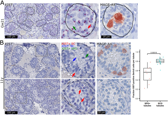
Klinefelter syndrome (47,XXY) causes infertility with a testicular histology comprising two types of Sertoli cell-only tubules, representing mature and immature-like Sertoli cells, and occasionally focal spermatogenesis. Here, we show that the immature-like Sertoli cells highly expressed XIST and had two X-chromosomes, while the mature Sertoli cells lacked XIST expression and had only one X-chromosome. Sertoli cells supporting focal spermatogenesis also lacked XIST expression and the additional X-chromosome, while the spermatogonia expressed XIST despite having only one X-chromosome. XIST was expressed in Sertoli cells until puberty, where a gradual loss was observed. Our results suggest that a micro-mosaic loss of the additional X-chromosome is needed for Sertoli cells to mature and to allow focal spermatogenesis.


Conflict of interest statement

The authors declare no competing interests.

Figures

Fig. 1
Fig. 1. XIST expression differentiates mature type A and immature-like type B tubules and is correlated with the number of X-chromosomes.
Sertoli cell-only (SCO) tubules classified as type A and type B on an HE staining were negative and positive for XIST RNA expression, respectively (A). Type A SCO tubules showed high protein levels of the Sertoli cell maturity markers androgen receptor (AR; B) and Clusterin (CLU; C), which was in contrast to type B tubules. DNA fluorescence in situ hybridization (FISH) against the X- (red) and Y-chromosome (green) (n = 1) revealed a tendency towards type A tubules being 46,XY and type B being 47,XXY. (D). DNA single-molecular (sm)ISH against chromosomes 3 (blue) and X (red) (n = 4) showed that type A tubules generally only contained one X-chromosome signal whereas type B tubules contained two (the tubules were identified on the basis of their nuclear morphology and XIST expression, see Fig. S6) (E, left). The mean fractions of Sertoli cells with two X-chromosome signals were around 15% in type A tubules (N = 31 tubules with a total of 887 Sertoli cells) and 69% in type B tubules (N = 23 tubules with a total of 927 Sertoli cells) and statistically significantly different between the two types of tubules (p = 4.7e-10, Wilcoxon Rank Sum Test). The fraction in type B tubules was similar to the one in Leydig cells (N = 20 areas with a total of 1291 Leydig cells) serving as an internal control (E, right). Scalebars represent 50 and 5 µm on the low and high magnifications, respectively.
Fig. 2
Fig. 2. XIST is expressed in spermatogonia but is absent in Sertoli cells supporting focal spermatogenesis, and both cells are euploid.
XIST-positive spermatogonia (SPG, yellow arrows) were present in testis specimens from men with Klinefelter syndrome, whereas the majority of the Sertoli cells (SC, white arrows) were XIST-negative (A). DNA FISH against the X- (red) and Y-chromosome (green) (n = 1) revealed a tendency towards spermatogonia and Sertoli cells supporting spermatogenesis being 46,XY (B). DNA smISH against chromosomes 3 (blue) and X (red) (n = 2) showed that spermatogonia and Sertoli cells generally only contained one X-chromosome signal (C). Scalebars represent 50 and 10 µm on the low and high magnifications, respectively. The mean fraction of spermatogonia (N = 19 tubules with 299 spermatogonia) and Sertoli cells (N = 13 tubules with 122 Sertoli cells) with two X-chromosome signals was around 15% and similar to type A tubules (lower shaded area) and very different from type B tubules and KS Leydig cells (upper shaded area) (D).
Fig. 3
Fig. 3. High XIST expression in Sertoli cells in fetuses but low XIST expression in tubules with spermatogonia compared to Sertoli cell-only (SCO) tubules in an 11-year-old prepubertal boy.
In a gestational week (Gw) 21 fetus, XIST was expressed in virtually all Sertoli cells (SC, red arrow) but showed a patchy expression pattern in spermatogonia (green arrow, identified with a MAGE-A4 antibody) (A). The same pattern was seen in three additional fetuses (Fig. S8). In an 11-year-old prepubertal boy, spermatogonia (SPG) also showed a patchy expression pattern of XIST (B, top; green arrow). In tubules with spermatogonia only a fraction of Sertoli cells expressed XIST (red arrow) while the remaining were negative (blue arrow). This was in contrast to SCO tubules (B, bottom). Scalebars represent 100 and 20 µm on the low and high magnifications, respectively. The mean fractions of XIST-positive Sertoli cells were around 52% in SPG+ tubules (N = 8 tubules with a total of 409 Sertoli cells) and 80% in SCO tubules (N = 16 tubules with 1373 Sertoli cells) and statistically significantly different between the two types of tubules (p = 0.00018, Wilcoxon Rank Sum Test) (B, right).
Fig. 4
Fig. 4. Schematic illustration of germ cell loss and the proposed model of X-chromosome loss in specific cell stages in the testes from men with Klinefelter syndrome (KS).
During fetal life (first column), the seminiferous tubules contain fetal germ cells (initially gonocytes and later pre-spermatogonia) and fetal Sertoli cells both in the 46,XY control (first row) and in KS (second and third row). In controls, the tubules contain spermatogonia and immature Sertoli cells in childhood (second column). Puberty leads to initiation of spermatogenesis (third column). For KS, testicular pathology is changing during childhood, puberty and adulthood leading to either a complete or partial loss of germ cells. Depending on the presence of germ cells and the ploidy of the Sertoli cells, four possible scenarios exist. The first three scenarios involve a complete loss of germ cells during childhood. In the first scenario, the Sertoli cells will attempt to mature leading to cell death and hyalinized tubules. In the second scenario, the Sertoli cells fail to mature thus creating morphologically distinct type B tubules. In the third scenario, a loss of the additional X-chromosome will lead to Sertoli cell maturation forming type A tubules. The fourth scenario implies that only a partial germ cell loss has occurred during childhood, and if the Sertoli cells and the spermatogonia lose the additional X-chromosome, focal spermatogenesis will occur. The figure is for illustration purposes and the actual order of events as well as the number of germ cells may be different. Figure created with BioRender.com.

References

    1. Jacobs PA, Strong JA. A case of human intersexuality having a possible XXY sex-determining mechanism. Nature. 1959;183:302–3. doi: 10.1038/183302a0. - DOI - PubMed
    1. Gravholt CH, Chang S, Wallentin M, Fedder J, Moore P, Skakkebæk A. Klinefelter syndrome: Integrating genetics, neuropsychology, and endocrinology. Endocr Rev. 2018;39:389–423. doi: 10.1210/er.2017-00212. - DOI - PubMed
    1. Berglund A, Viuff MH, Skakkebæk A, Chang S, Stochholm K, Gravholt CH. Changes in the cohort composition of turner syndrome and severe non-diagnosis of Klinefelter, 47,XXX and 47,XYY syndrome: A nationwide cohort study 11 Medical and Health Sciences 1117 Public Health and Health Services. Orphanet J Rare Dis. 2019;14:1–9. - PMC - PubMed
    1. Herlihy AS, Halliday JL, Cock ML, McLachlan RI. The prevalence and diagnosis rates of Klinefelter syndrome: An Australian comparison. Med J Aust. 2011;194:24–8. doi: 10.5694/j.1326-5377.2011.tb04141.x. - DOI - PubMed
    1. Bojesen A, Juul S, Gravholt CH. Prenatal and postnatal prevalence of Klinefelter syndrome: A national registry study. J Clin Endocrinol Metab. 2003;88:622–6. doi: 10.1210/jc.2002-021491. - DOI - PubMed

Publication types