. 2024 Jun 5;8(1):127.
doi: 10.1038/s41698-024-00593-y.

Blocking Oncostatin M receptor abrogates STAT3 mediated integrin signaling and overcomes chemoresistance in ovarian cancer

Affiliations

Blocking Oncostatin M receptor abrogates STAT3 mediated integrin signaling and overcomes chemoresistance in ovarian cancer

Anjali Geethadevi et al. NPJ Precis Oncol. .

Abstract

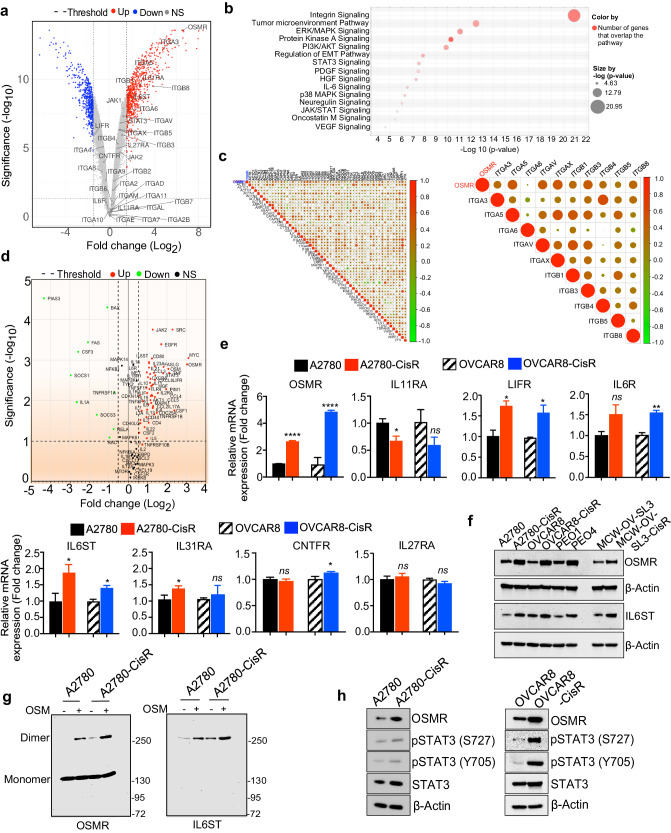
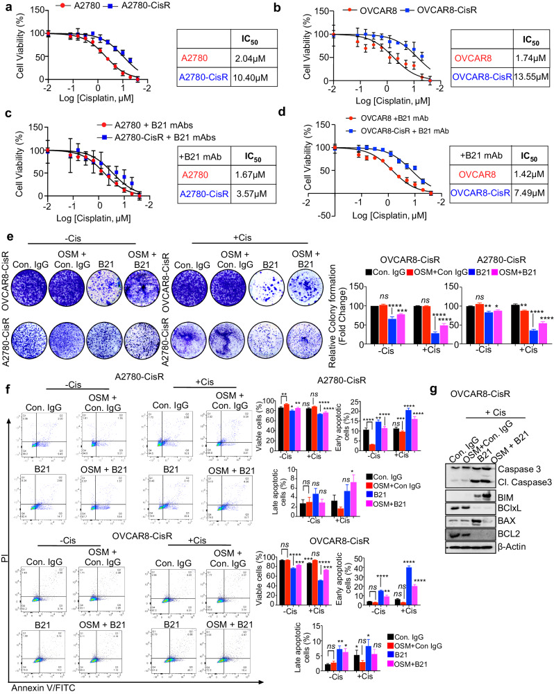
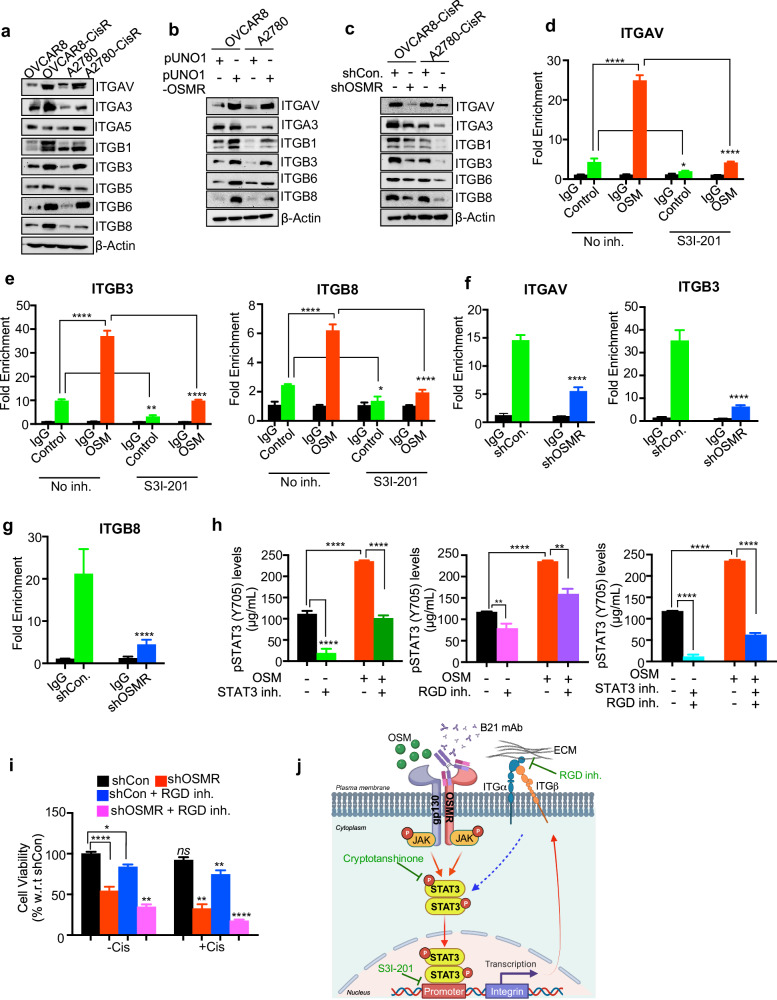
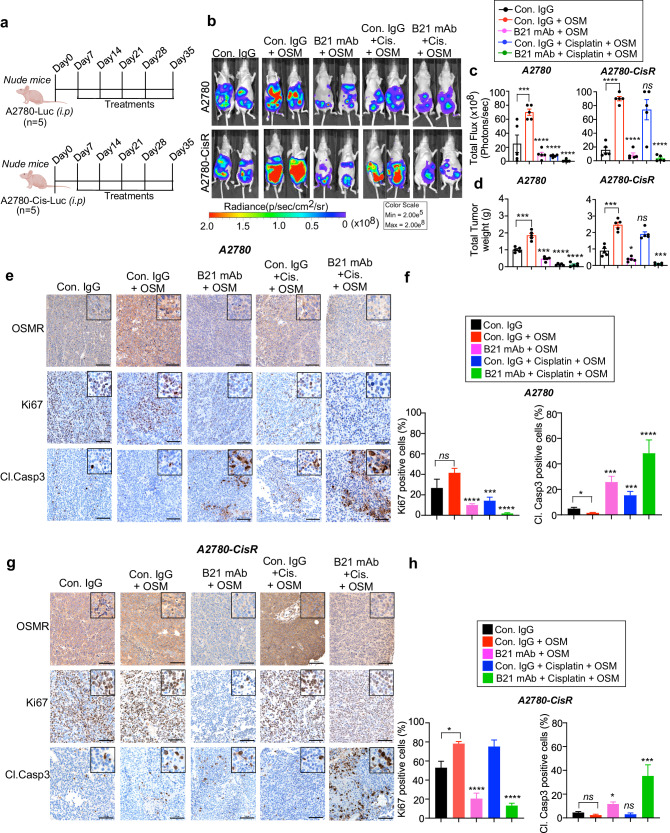
Chemotherapy such as cisplatin is widely used to treat ovarian cancer either before or after surgical debulking. However, cancer relapse due to chemotherapy resistance is a major challenge in the treatment of ovarian cancer. The underlying mechanisms related to chemotherapy resistance remain largely unclear. Therefore, identification of effective therapeutic strategies is urgently needed to overcome therapy resistance. Transcriptome-based analysis, in vitro studies and functional assays identified that cisplatin-resistant ovarian cancer cells express high levels of OSMR compared to cisplatin sensitive cells. Furthermore, OSMR expression associated with a module of integrin family genes and predominantly linked with integrin αV (ITGAV) and integrin β3 (ITGB3) for cisplatin resistance. Using ectopic expression and knockdown approaches, we proved that OSMR directly regulates ITGAV and ITGB3 gene expression through STAT3 activation. Notably, targeting OSMR using anti-OSMR human antibody inhibited the growth and metastasis of ovarian cancer cells and sensitized cisplatin treatment. Taken together, our results underscore the pivotal role of OSMR as a requirement for cisplatin resistance in ovarian cancer. Notably, OSMR fostered the expression of a distinct set of integrin genes, which in turn resulted into a crosstalk between OSMR and integrins for signaling activation that is critical for cisplatin resistance. Therefore, targeting OSMR emerges as a promising and viable strategy to reverse cisplatin-resistance in ovarian cancer.


Conflict of interest statement

P.C.-R, S.P., A.G., Z.K., N.Z., and Z.A. are inventors of a patent PCT/US2022/074276 on OSMR antibody discovery and therapeutic uses. The other authors have no competing interests to declare.

Figures

Fig. 1
Fig. 1. Oncostatin M (OSM) receptor is upregulated in chemoresistant ovarian cancer through OSMR heterodimerization.
a Volcano plot shows differentially expressed genes in OVCAR8-CisR (cisplatin resistant) spheroids (N = 4) compared with OVCAR8 sensitive spheroids (N = 4) in GSE45553 microarray dataset. Significance threshold is P ≤ 0.05 and Log2 (fold change) threshold set at ±0.585 on both sides. Top hits and OSM module genes are labeled (P value < 0.05, FDR < 0.1). ns non-significant. b Ingenuity Pathway Analysis (IPA) shows canonical pathways that are >1.5-fold change in OVCAR8-CisR spheroids (GSE45553) with a threshold cut off log10 (fold change) > 4 respectively. c Correlation graph shows Spearman’s correlation of OSMR with (left panel) Integrin family genes and (right panel) with integrins α and β genes exhibiting P value ≤ 0.05 in TCGA ovarian cancer cohort (n = 379) dataset downloaded from GDC data portal. d Volcano plot shows differentially expressed genes in A2780 CisR cell line compared with A2780 sensitive cell lines in triplicates in the qPCR array panel of IL6 signaling. Genes were selected based on P value ≤ 0.05 and Log2 (fold change) threshold set at +/−0.58 in both directions. ns: non-significant. e Total RNA was isolated from the indicated cell lines and mRNA expression of IL6 receptor family genes were determined. β-actin gene was used as an internal standard. f Western blot shows OSMR and IL6ST expression in the lysates of a set of cisplatin-sensitive and resistant ovarian cancer cell lines. g OSMR was immunoprecipitated (IP) from A2780-CisR and A2780 sensitive cells were treated with recombinant human OSM (100 ng/mL) and crosslinked with BS3 agent. Monomers and dimers obtained by IP were resolved on SDS-PAGE and immunoblotted using OSMR and IL6ST specific antibodies. h Western blot shows the level of OSMR, and total and phosphorylated form of STAT3 in both cisplatin sensitive and resistant version of A2780 and OVCAR8 cells. Student’s t test (two tailed, unpaired) was performed to determine significance between the groups. Error bars represent mean ± SEM. ****P ≤ 0.0001, **P ≤ 0.01, *P ≤ 0.05, ns non-significant.
Fig. 2
Fig. 2. OSMR overexpression promotes chemoresistance.
a, b Cell viability was determined using CCK8 reagent in both control and OSMR overexpressing OVCAR8 and A2780 cells were treated with different concentrations of cisplatin for 48 h. The IC50 of cisplatin in both control and OSMR overexpressing cell lines were quantitated and presented below in the respective boxes. c, d OVCAR8 and A2780 cells stably overexpressing OSMR, and the control cells were lysed and immunoblotted using indicated antibodies. e, f mRNA expression of cancer stemness markers and EMT markers were evaluated using qPCR in OVCAR8 and A2780 cells stably overexpressing OSMR and the control cells. g OVCAR8 and A2780 cells stably overexpressing OSMR and the control cells were grown in the presence and absence of cisplatin 5 μM for 15 days and imaged (left panel). Colonies formed were lysed using 10% acetic acid and absorbance were quantitated at 560 nm (right panel). h Control and OSMR overexpressing OVCAR8 and A2780 cells were treated with and without cisplatin for 16 h and the level of cellular apoptosis was quantitated using Annexin V-APC/7-AAD staining followed by flow cytometry (left). Bar graph shows percentage of viable cells, cells undergoing early, or late apoptosis quantitated from flow cytometry. Individual data points are represented as mean ± SEM. Student’s t test (two tailed, unpaired) was performed to determine significance between groups. ****P ≤ 0.0001, ***P ≤ 0.001, **P ≤ 0.01, *P ≤ 0.05, ns non-significant.
Fig. 3
Fig. 3. Anti-OSMR antibody enhances sensitivity towards cisplatin.
a, b Cell viability was measured using CCK8 reagent in A2780 versus A2780-CisR cells, OVCAR8 versus OVCAR8-CisR cells were treated with different concentrations of cisplatin for 48 h. The IC50 of cisplatin in both control and OSMR overexpressing cell lines were quantitated and presented below in the respective boxes. c, d Cell viability assay was measured using CCK8 reagent in A2780 and A2780-CisR and OVCAR8 vs. OVCAR8-CisR resistant cells treated with anti-OSMR B21 mAb (10 μg/mL) in combination with the indicated concentrations of cisplatin for 48 h. The IC50 of B21 mAb in both control and OSMR overexpressing cell lines were quantitated and presented below in the respective boxes. e OVCAR8-CisR and A2780-CisR cells were grown in the presence and absence of B21 antibody or isotype control IgG (10 μg/mL each) with or without recombinant human rhOSM (100 ng/mL) for 15 days in 6-well plate. Colonies formed were stained using 0.5% of crystal violet and imaged. Colonies formed were lysed using 10% acetic acid and absorbance were quantitated at 560 nm (right panel). f Annexin V-FITC/PI based apoptosis assay was performed using flow cytometry in OVCAR8-CisR and A2780-CisR resistant cells that were treated with B21 antibody or isotype control IgG (10 μg/mL each) in combination with/without cisplatin and stimulated in the presence and absence of rhOSM (100 ng/mL) for 16 h. The representative quantitation histograms show percentage of cells that are viable and undergoing early or late apoptosis. g Western blot shows the expression of pro-apoptotic and anti-apoptotic proteins in OVCAR8-CisR cell lines that were treated with B21 anti-OSMR antibodies (10 µg/mL) in combination with cisplatin in the presence and absence of rhOSM (100 ng/mL) for 24 h. One-way ANOVA with Dunnett’s multiple comparison test was performed for significance. Data represent means ± SEM. ****P ≤ 0.0001, ***P ≤ 0.001, **P ≤ 0.01, *P ≤ 0.05, ns non-significant.
Fig. 4
Fig. 4. OSMR transcriptionally regulates integrins via STAT3 which facilitates OSMR signaling in promoting chemoresistance.
a Western blot analysis shows the levels of integrins in OVCAR8, OVCAR8-CisR, A2780, A2780-CisR cells. b Western blot shows the level of selected integrins in both OVCAR8 and A2780 control cells and the cells stably overexpress OSMR. c OSMR was stably knocked down in cisplatin resistant OVCAR8 and A2780 cells and Western blot was performed using indicated antibodies specific to integrins. d, e OVCAR8 cells were stimulated in the presence and absence of rhOSM (100 ng/mL), with or without STAT3 inhibitor S3I-201 (50 μM) for 48 h. Chromatin-immunoprecipitation (ChIP) was performed using STAT3 antibody and qPCR was performed to determine the enrichment of promoters of ITGAV, ITGB3 and ITGB8 genes. The qPCR data are presented as fold enrichment compared to IgG control. f, g OSMR knocked down OVCAR8-CisR cells and empty vector expressing cells (shControl) were lysed, and chromatin-immunoprecipitation was performed using STAT3 antibody. Promoter enrichment of indicated genes were quantitated from the samples. h OVCAR8 cells were seeded on FN-precoated 96-well plates and treated with either STAT3 inhibitor, RGD peptide inhibitor or both for 30 min with or without rhOSM. Cells were lysed and pSTAT3 (Y705) levels were quantitated using ELISA. i OVCAR8-CisR cells stably knockdown with shOSMR were treated with RGD peptide inhibitor in the presence and absence of cisplatin for 48 h and cell viability assay was determined using CCK8. j Schema demonstrates how OSMR upregulates integrins through STAT3 activation and how does blocking of OSMR abrogates STAT3 mediated integrin levels. For significance, one-way ANOVA followed by Dunnett’s multiple comparison test were performed in ‘h’ and ‘i’ and Student’s t test (two tailed, unpaired) was performed for significance in ‘d, e’. Error bars represent means ± SEM. ****P ≤ 0.0001, ***P ≤ 0.001, **P ≤ 0.01, *P ≤ 0.05, ns: non-significant.
Fig. 5
Fig. 5. Anti-OSMR antibody increases the chemosensitivity of ovarian cancer cells towards Cisplatin in vivo.
a Schema shows how A2780-Luc+ and A2780-CisR-Luc+ cells were grown in athymic nude mice and treated with isotype control IgG or B21 mAb (10 mg/kg body weight; i.p.) in the presence and absence of Cisplatin (5 mg/kg body weight) for 5 weeks. Mice were treated with OSM (250 ng/kg body weight) 30 minutes after the injections of control IgG, or B21 antibodies (10 mg/kg body weight) i.p. b Representative images showing rate of tumor growth was determined by bioluminescence imaging (BLI) using IVIS100 imager. c Quantitative assessment of luciferase signal intensity of A2780-Luc+ and A2780-CisR-Luc+ tumor bearing mice following the treatments before termination on day 35. d Total weight of the tumor in the respective treatment groups were quantitated. e, g Tumor tissues from A2780 and A2780-CisR bearing mice were subjected to immunohistochemistry using the indicated antibodies. Whole stained tissues sections were scanned, and representative images of the indicated proteins from each group were captured in ×20. Scale bar = 100 μm. f, h Quantitative assessment of Ki67 and cleaved caspase 3 cells in the respective treatment groups. One-way ANOVA with Dunnett’s multiple comparison test or Student’s t test were performed for determining significance. Data represent means ± SEM. ****P ≤ 0.0001, ***P ≤ 0.001, **P ≤ 0.01, *P ≤ 0.05. ns: non-significant.

References

    1. Marcus CS, Maxwell GL, Darcy KM, Hamilton CA, McGuire WP. Current approaches and challenges in managing and monitoring treatment response in ovarian cancer. J. Cancer. 2014;5:25–30. doi: 10.7150/jca.7810. - DOI - PMC - PubMed
    1. Wang Y, et al. Autocrine production of interleukin-6 confers cisplatin and paclitaxel resistance in ovarian cancer cells. Cancer Lett. 2010;295:110–123. doi: 10.1016/j.canlet.2010.02.019. - DOI - PubMed
    1. Wang, Y. et al. IL-6 mediates platinum-induced enrichment of ovarian cancer stem cells. JCI Insight10.1172/jci.insight.122360 (2018). - PMC - PubMed
    1. Masjedi A, et al. Oncostatin M: A mysterious cytokine in cancers. Int. Immunopharmacol. 2021;90:107158. doi: 10.1016/j.intimp.2020.107158. - DOI - PubMed
    1. Parashar D, et al. miRNA551b-3p activates an oncostatin signaling module for the progression of triple-negative breast cancer. Cell Rep. 2019;29:4389–4406.e4310. doi: 10.1016/j.celrep.2019.11.085. - DOI - PMC - PubMed