. 2024 Jul;45(7):1425-1437.
doi: 10.1038/s41401-024-01306-8. Epub 2024 Jun 5.

Acetylcytidine modification of Amotl1 by N-acetyltransferase 10 contributes to cardiac fibrotic expansion in mice after myocardial infarction

Affiliations

Acetylcytidine modification of Amotl1 by N-acetyltransferase 10 contributes to cardiac fibrotic expansion in mice after myocardial infarction

Xiu-Xiu Wang et al. Acta Pharmacol Sin. 2024 Jul.

Abstract

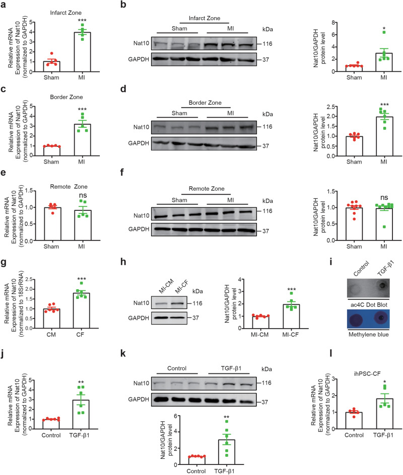
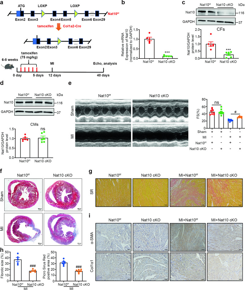
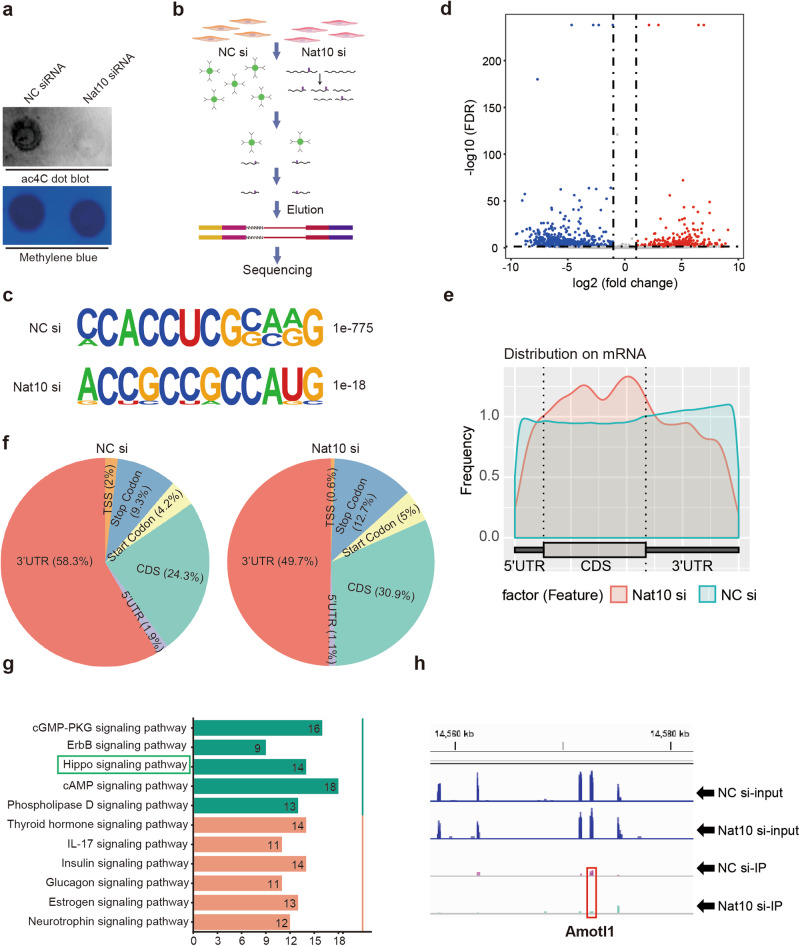
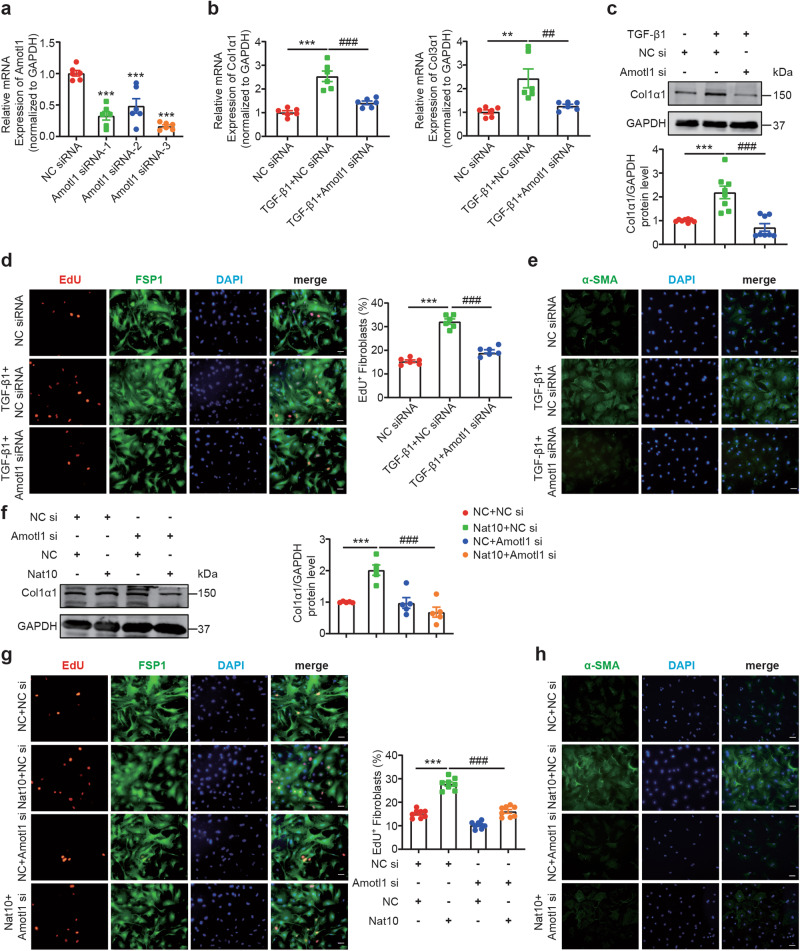
Cardiac fibrosis is a pathological scarring process that impairs cardiac function. N-acetyltransferase 10 (Nat10) is recently identified as the key enzyme for the N4-acetylcytidine (ac4C) modification of mRNAs. In this study, we investigated the role of Nat10 in cardiac fibrosis following myocardial infarction (MI) and the related mechanisms. MI was induced in mice by ligation of the left anterior descending coronary artery; cardiac function was assessed with echocardiography. We showed that both the mRNA and protein expression levels of Nat10 were significantly increased in the infarct zone and border zone 4 weeks post-MI, and the expression of Nat10 in cardiac fibroblasts was significantly higher compared with that in cardiomyocytes after MI. Fibroblast-specific overexpression of Nat10 promoted collagen deposition and induced cardiac systolic dysfunction post-MI in mice. Conversely, fibroblast-specific knockout of Nat10 markedly relieved cardiac function impairment and extracellular matrix remodeling following MI. We then conducted ac4C-RNA binding protein immunoprecipitation-sequencing (RIP-seq) in cardiac fibroblasts transfected with Nat10 siRNA, and revealed that angiomotin-like 1 (Amotl1), an upstream regulator of the Hippo signaling pathway, was the target gene of Nat10. We demonstrated that Nat10-mediated ac4C modification of Amotl1 increased its mRNA stability and translation in neonatal cardiac fibroblasts, thereby increasing the interaction of Amotl1 with yes-associated protein 1 (Yap) and facilitating Yap translocation into the nucleus. Intriguingly, silencing of Amotl1 or Yap, as well as treatment with verteporfin, a selective and potent Yap inhibitor, attenuated the Nat10 overexpression-induced proliferation of cardiac fibroblasts and prevented their differentiation into myofibroblasts in vitro. In conclusion, this study highlights Nat10 as a crucial regulator of myocardial fibrosis following MI injury through ac4C modification of upstream activators within the Hippo/Yap signaling pathway.

Keywords: Hippo signaling pathway; N-acetyltransferase 10; ac4C-RIP-sequencing; angiomotin-like 1; cardiac fibrosis.


Conflict of interest statement

The authors declare no competing interests.

Figures

Fig. 1
Fig. 1. Upregulation of Nat10 expression in mouse hearts after MI.
af qRT-PCR and Western blot analyses of alterations in the Nat10 mRNA and protein levels in the infarct, border and remote zones of cardiac tissues 28 days post MI compared to those in the Sham group. GAPDH was used as a loading control. g The Nat10 mRNA levels in cardiomyocytes and fibroblasts isolated from the left ventricles of adult mice (n = 6) were measured by qRT-PCR. 18 S rRNA was used as an internal reference. h Representative images of immunoblots showing the Nat10 protein level in cardiac fibroblasts and cardiomyocytes at 4 weeks after MI (n = 6). Furthermore, neonatal cardiac fibroblasts were isolated and treated with TGF-β1 (10 ng/ml) for 24 h, and i a dot blot assay was performed to assess ac4C modification, j qRT-PCR was used to measure Nat10 mRNA expression, and k representative immunoblotting images were acquired to show the Nat10 protein level upon TGF-β1-mediated induction of the fibrotic response. In addition, the change in the Nat10 mRNA level in TGF-β1-induced (24 h) pluripotent stem cell-derived cardiac fibroblasts was evaluated by qRT-PCR l. An unpaired t-test was used for comparisons between the two groups. All the data are presented as the means ± SEMs. *P < 0.05, **P < 0.01, ***P < 0.001 versus the Sham, cardiomyocyte or Control group.
Fig. 2
Fig. 2. Fibroblast-specific overexpression of Nat10 results in cardiac fibrosis and aggravates myocardial fibrosis after MI.
a Schematic diagram of the process for fibroblast-specific overexpression of Nat10. Fibroblasts were isolated from Nat10; Col1a2-CreER mice and their wild-type littermates, and b the efficiency of Nat10 knockout in transgenic mice was determined by qRT-PCR. n = 5. c Representative images of immunoblots showing the Nat10 protein levels in fibroblasts from Nat10 transgenic mice (Nat10; Col1a2-CreER, n = 6) and their wild-type (WT, n = 6) littermates. Moreover, d the levels of Nat10 in cardiomyocytes isolated from Nat10; Col1a2-CreER mice and their wild-type littermates were measured by Western blotting. n = 6. e FS was calculated 4 weeks after MI. f, g The fibrotic area in Nat10 transgenic mice was assessed by Masson’s trichrome staining, collagen deposition was evaluated via picrosirius red staining and the results were compared to those in the MI + WT group. In addition, adult cardiac fibroblasts were isolated and transfected with the Nat10 plasmid. After 48 h, representative images of immunoblots showing the Col1α1 and its protein level were obtained h. FS, Fractional Shortening. An unpaired t-test was used for comparisons between the two groups. All the data are presented as the means ± SEMs. *P < 0.05, **P < 0.01, ***P < 0.001 versus the WT or NC groups, #P < 0.05, ###P < 0.001 versus the MI + WT group.
Fig. 3
Fig. 3. Fibroblast-specific Nat10 knockout attenuates cardiac fibrosis after MI.
a Schematic diagram of the process for Col1a2-specific deletion in Nat10 mice and the subsequent experimental procedure. b The efficiency of Nat10 knockdown was determined by qRT‒PCR in isolated fibroblasts. n = 5. Representative images of immunoblots and quantification of the Nat10 protein level are shown for cardiac fibroblasts c and cardiomyocytes d isolated from Nat10f/f and Nat10 cKO mice. e Cardiac function was evaluated by echocardiography in the Nat10f/f, Nat10 cKO, and MI+ Nat10f/f, MI + Nat10 cKO groups 4 weeks after MI. fh Effect of Nat10 ablation on scar size and collagen deposition at detected 4 weeks after MI. Scale bars: 500 µm. i In the 4 groups, α-SMA+ or Col1α1+ cells were detected by immunohistochemistry. n = 6. Scale bars: 50 µm. All the data are presented as the means ± SEMs. ***P < 0.001 versus the Nat10f/f group, #P < 0.05, ###P < 0.001 versus the MI + Nat10f/f group.
Fig. 4
Fig. 4. Screening for Nat10-related differentially expressed genes by ac4C-RIP followed by sequencing.
a A dot Blot assay was performed to detect ac4C modification after knockdown of Nat10. Methylene blue staining was used as an internal reference for a consistent sample size. b Schematic illustration of the process for ac4C-RIP-seq of cardiac fibroblasts transfected with NC siRNA and Nat10 siRNA. c Enriched motifs of ac4C peaks were identified in the two groups. d The differentially expressed ac4C-modified genes between the two groups are shown in the volcano plot. e Metagene plot and f pie chart displaying the distribution of ac4C modifications within the acetylated mRNAs. g KEGG signaling pathway enrichment analysis of the differentially expressed ac4C-modified mRNAs. h Ac4C peak on the Amotl1 mRNA.
Fig. 5
Fig. 5. Identification of Amotl1 as a target of Nat10 and its dependency on Nat10-mediated ac4C modification.
Cardiac fibroblasts were isolated from neonatal mice (1-3 days old) and transfected with the Nat10 plasmid or siRNA for 48 h. a, b The abundance of ac4C in the Amotl1 mRNA upon Nat10 overexpression and knockdown was measured by acRIP-qPCR. c, d The effect of actinomycin D on Amotl1 stability was assessed at 0, 2, 4, and 6 h post-treatment using qRT-PCR. e, f The impact of actinomycin D on Amotl1 translation was evaluated via Western blot analysis. Cardiac fibroblasts were transfected with the NC or Nat10 mutant (Mut-641) plasmid, and after 48 h, g the ac4C modification of Amotl1 was detected via ac4C-RIP-PCR. h, i Furthermore, the protein level of the Yap protein in the cytoplasm and nucleus upon Nat10 overexpression or knockdown was assessed. Simultaneously, immunofluorescence staining (Yap labeled in green) was employed to demonstrate the subcellular localization of Yap in the cytoplasm and nucleus j, k. Finally, Western blot analysis was carried out to assess the impact of Nat10 Mut expression on the Yap level in the cytoplasm and nucleus l. Scale bars: 50 µm. All the data are presented as the means ± SEMs. *P < 0.05, **P < 0.01, ***P < 0.001 versus the NC-IP, NC, NC + DMSO or NC siRNA groups, #P < 0.05 versus the NC + ActD or NC siRNA + ActD groups.
Fig. 6
Fig. 6. Silencing of Amotl1 significantly inhibits the TGF-β1-mediated proliferation and transdifferentiation of fibroblasts and the Nat10-induced fibrotic response.
Following the transfection of Amotl1 siRNA into isolated mouse fibroblasts for 48 h, a the efficiency of Amotl1 knockdown was assessed using qRT-PCR. b Inhibiting Amotl1 expression reduced the mRNA levels of Col1α1 and Col3α1, as well as the protein level of Col1α1 c. d Silencing Amotl1 significantly inhibited fibroblast proliferation, as determined via an EdU incorporation assay (n = 6), and suppressed the transdifferentiation of fibroblasts into myofibroblasts in the TGF-β1-mediated fibrotic response e. Furthermore, after cotransfection of the Nat10 plasmid and Amotl1 siRNA into cardiac fibroblasts for 48 h, f the protein level of Col1α1 was measured by Western blotting. g Fibroblast proliferation was assessed using an EdU assay (n = 8). h Fibroblast transdifferentiation into myofibroblasts was evaluated via an immunofluorescence assay with α-SMA (green) staining. All the data are presented as the means ± SEMs. **P < 0.01, ***P < 0.001 versus the NC siRNA or NC siRNA + NC groups, ##P < 0.01, ###P < 0.001 versus the TGF-β1 + NC siRNA or Nat10 + NC siRNA groups.
Fig. 7
Fig. 7. Silencing of Yap or verteporfin blocks the fibrotic response induced by Nat10 overexpression.
Cells were co-transfected with the Nat10 plasmid and the Yap siRNA for 48 h, a qRT-PCR was used to measure the mRNA expression of Col1α1 and Col3α1, while the protein level of Col1α1 was assessed by Western blotting b. Moreover, an EdU assay (n = 8) was employed to evaluate the effect of silencing Yap on Nat10-mediated fibroblast proliferation c. After transfection with the Nat10 overexpression plasmid for 24 h, 1 μM verteporfin (Yap inhibitor) was added for an additional 24 h, d the Col1α1 and Col3α1 mRNA levels and the Col1α1 protein level e were measured. f An EdU incorporation assay (n = 7) was performed to assess fibroblast proliferation and differentiation into myofibroblasts was evaluated via immunofluorescence assay with α-SMA (green) staining g. Scale bars: 50 µm. All the data are presented as the means ± SEMs. **P < 0.01, ***P < 0.001 versus the NC + NC siRNA or NC + DMSO group, #P < 0.05, ##P < 0.01, ###P < 0.001 versus the Nat10 + NC siRNA or Nat10 + DMSO group.
Fig. 8
Fig. 8. Targeting Nat10 alleviates the TGF-β1-mediated fibrotic response in human cardiac fibroblasts in vitro.
A human cardiac fibroblast line was transfected with the Nat10 siRNA after the addition of TGF-β1 (10 ng/ml), and after 48 h, a the evaluation of mRNA expression levels of Col1α1 and Col3α1 were measured. b Knockdown of Nat10 affected the proliferation of human fibroblast cell lines as observed by EdU staining. n = 6. Scale bars: 50 µm. All the data are presented as the means ± SEMs. ***P < 0.001 versus the NC siRNA group, #P < 0.05, ###P < 0.001 versus the TGF-β1 + NC siRNA group.

Similar articles

Cited by

References

    1. Tallquist MD. Cardiac fibroblast diversity. Annu Rev Physiol. 2020;82:63–78. doi: 10.1146/annurev-physiol-021119-034527. - DOI - PMC - PubMed
    1. Travers JG, Kamal FA, Robbins J, Yutzey KE, Blaxall BC. Cardiac fibrosis: the fibroblast awakens. Circ Res. 2016;118:1021–40. doi: 10.1161/CIRCRESAHA.115.306565. - DOI - PMC - PubMed
    1. Pinto AR. Matricellular proteins as critical regulators of fibrosis. Circ Res. 2021;129:1036–8. doi: 10.1161/CIRCRESAHA.121.320273. - DOI - PubMed
    1. Stratton MS, Bagchi RA, Felisbino MB, Hirsch RA, Smith HE, Riching AS, et al. Dynamic chromatin targeting of BRD4 stimulates cardiac fibroblast activation. Circ Res. 2019;125:662–77. doi: 10.1161/CIRCRESAHA.119.315125. - DOI - PMC - PubMed
    1. Zhang QJ, Tran TAT, Wang M, Ranek MJ, Kokkonen-Simon KM, Gao J, et al. Histone lysine dimethyl-demethylase KDM3A controls pathological cardiac hypertrophy and fibrosis. Nat Commun. 2018;9:5230. doi: 10.1038/s41467-018-07173-2. - DOI - PMC - PubMed

MeSH terms