. 2024 Jun 5;21(1):148.
doi: 10.1186/s12974-024-03143-2.

CD36 deletion prevents white matter injury by modulating microglia polarization through the Traf5-MAPK signal pathway

Affiliations

CD36 deletion prevents white matter injury by modulating microglia polarization through the Traf5-MAPK signal pathway

Xiaoxiang Hou et al. J Neuroinflammation. .

Abstract

Background: White matter injury (WMI) represents a significant etiological factor contributing to neurological impairment subsequent to Traumatic Brain Injury (TBI). CD36 receptors are recognized as pivotal participants in the pathogenesis of neurological disorders, including stroke and spinal cord injury. Furthermore, dynamic fluctuations in the phenotypic polarization of microglial cells have been intimately associated with the regenerative processes within the injured tissue following TBI. Nevertheless, there is a paucity of research addressing the impact of CD36 receptors on WMI and microglial polarization. This investigation aims to elucidate the functional role and mechanistic underpinnings of CD36 in modulating microglial polarization and WMI following TBI.

Methods: TBI models were induced in murine subjects via controlled cortical impact (CCI). The spatiotemporal patterns of CD36 expression were examined through quantitative polymerase chain reaction (qPCR), Western blot analysis, and immunofluorescence staining. The extent of white matter injury was assessed via transmission electron microscopy, Luxol Fast Blue (LFB) staining, and immunofluorescence staining. Transcriptome sequencing was employed to dissect the molecular mechanisms underlying CD36 down-regulation and its influence on white matter damage. Microglial polarization status was ascertained using qPCR, Western blot analysis, and immunofluorescence staining. In vitro, a Transwell co-culture system was employed to investigate the impact of CD36-dependent microglial polarization on oligodendrocytes subjected to oxygen-glucose deprivation (OGD).

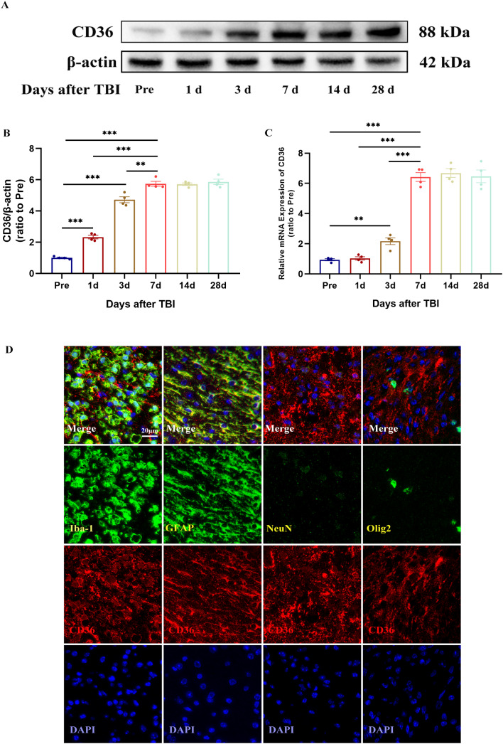
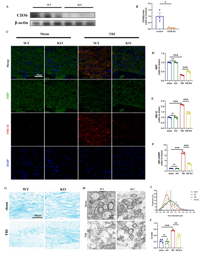
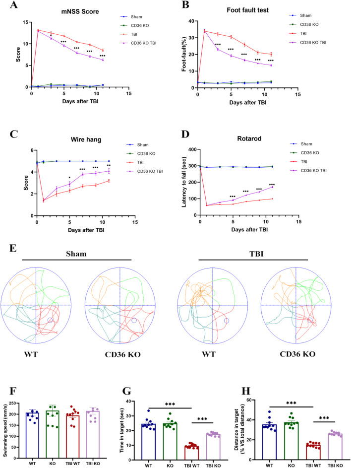
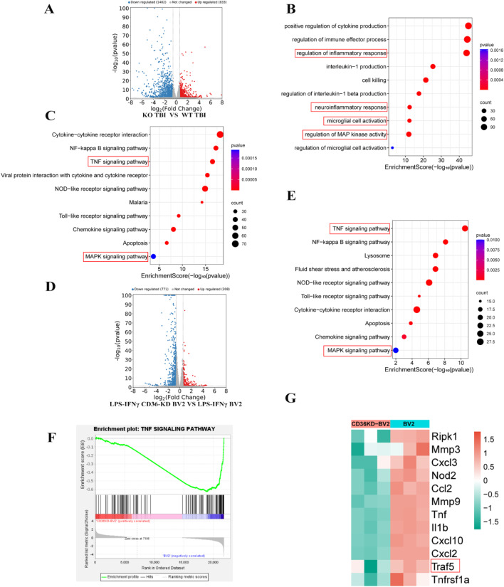
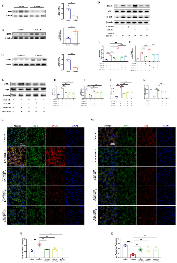
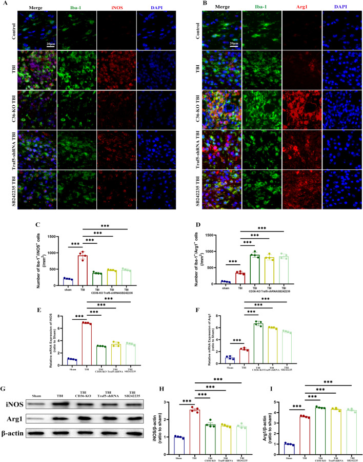
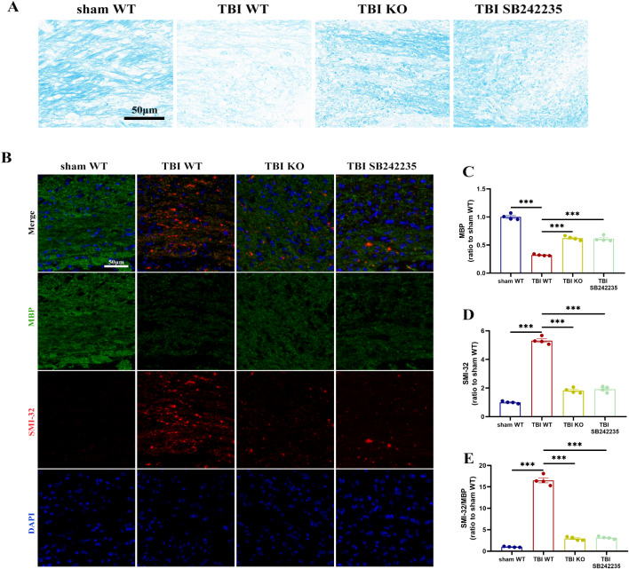
Results: Western blot and qPCR analyses revealed that CD36 expression reached its zenith at 7 days post-TBI and remained sustained at this level thereafter. Immunofluorescence staining exhibited robust CD36 expression in astrocytes and microglia following TBI. Genetic deletion of CD36 ameliorated TBI-induced white matter injury, as evidenced by a reduced SMI-32/MBP ratio and G-ratio. Transcriptome sequencing unveiled differentially expressed genes enriched in processes linked to microglial activation, regulation of neuroinflammation, and the TNF signaling pathway. Additionally, bioinformatics analysis pinpointed the Traf5-p38 axis as a critical signaling pathway. In vivo and in vitro experiments indicated that inhibition of the CD36-Traf5-MAPK axis curtailed microglial polarization toward the pro-inflammatory phenotype. In a Transwell co-culture system, BV2 cells treated with LPS + IFN-γ exacerbated the damage of post-OGD oligodendrocytes, which could be rectified through CD36 knockdown in BV2 cells.

Conclusions: This study illuminates that the suppression of CD36 mitigates WMI by constraining microglial polarization towards the pro-inflammatory phenotype through the down-regulation of the Traf5-MAPK signaling pathway. Our findings present a potential therapeutic strategy for averting neuroinflammatory responses and ensuing WMI damage resulting from TBI.

Keywords: CD36; Microglial polarization; Traumatic brain injury; White matter injury.


Conflict of interest statement

The authors declare that they have no competing interests.

Figures

Sch. 1
Sch. 1
Schematic diagram of time points for experiments of this study. CCI: controlled cortical impact; OGD: oxygen-glucose deprivation
Fig. 1
Fig. 1
Temporal and spatial expression patterns of CD36 receptor expression after TBI. (A, B) Western blot analysis and quantification depicting the temporal expression of CD36 receptors in brain tissue post-TBI. (C) Quantitative PCR results showing CD36 expression trends following TBI. (D) Immunofluorescence images illustrating CD36 localization in neurons, microglia, astrocytes, and oligodendrocytes at day 7 post-injury within the injury and peri-injury zones. Scale bar: 20 μm. Data represent mean ± SEM for 4 mice per group. *P < 0.05, **P < 0.01, ***P < 0.001
Fig. 2
Fig. 2
CD36 knockout mitigates white matter injury. (A, B) Validation of CD36 knockout efficiency via western blot and associated quantification, n = 3/group. (C) Luxol fast blue staining demonstrating reduced myelin loss in the corpus callosum of CD36 knockout mice at day 7 post-TBI. Scale bar: 50 μm. (D) Transmission electron microscopy images showing demyelination in the corpus callosum at day 7 post-TBI. Scale bar: 1 μm. (E) Axonal diameter distribution. (F) G-ratio analysis reflecting myelin thickness relative to axonal diameter. (G) Dual immunofluorescence staining for dephosphorylated neurofilament protein (SMI-32, red) and myelin basic protein (MBP, green) in the corpus callosum at day 7 post-TBI, with DAPI staining nuclei (blue). Scale bar: 50 μm. (HJ) Quantitative analysis of white matter injury, indicated by changes in MBP (H), SMI-32 (I), and their ratio (J). Data represent mean ± SEM for 4 mice per group. *P < 0.05, **P < 0.01, ***P < 0.001
Fig. 3
Fig. 3
Neurological function enhancement in CD36 knockout mice. (A) Modified Neurological Severity Scores (mNSS) over time post-TBI. (BD) Sensorimotor assessments including the foot fault, wire hang, and rotarod tests. (EH) Cognitive function evaluation in the Morris water maze, with swim paths depicted during the probe trial (E), distance traveled (H), and time spent in the target quadrant (G), without differences in swim speed (F). Data represent mean ± SEM for 10 mice per group. *P < 0.05, **P < 0.01, ***P < 0.001
Fig. 4
Fig. 4
Transcriptomic analysis post-CD36 inhibition. (A) Volcano plot illustrating differentially expressed genes (DEGs) between CD36(-/-) and wild-type mice post-TBI. (B) Gene Ontology (GO) enrichment analysis highlighting biological processes among DEGs. (C) Kyoto Encyclopedia of Genes and Genomes (KEGG) pathway enrichment analysis of DEGs. (D) Volcano plot of DEGs between CD36-knockdown (KD) BV2 cells and control BV2 cells treated with LPS + IFN-γ. (E) KEGG pathway enrichment analysis for DEGs in CD36-KD BV2 cells. (F) Gene Set Enrichment Analysis (GSEA) for the TNF signaling pathway. (G) Identification of core genes via GSEA.
Fig. 5
Fig. 5
CD36 knockdown reduces pro-inflammatory polarization in BV2 cells via the Traf5-MAPK axis. (AC) Validation of stable transfection in BV2 cells by western blot and quantification, n = 3/group. (DF) Assessment of the Traf5-MAPK signaling pathway by western blot and quantification. (GI) Western blot analysis of iNOS and ARG1 expression. (J, K) qPCR validation of iNOS and ARG1 expression. (L, M) Immunofluorescence images depicting pro-inflammatory (L) and anti-inflammatory (M) polarization in BV2 cells across different experimental conditions. (N, O) Proportional analysis of polarized BV2 cells relative to the total BV2 population. Pro-inflammatory phenotype/Total (N); Anti-inflammatory phenotype/Total (O). Data represent mean ± SEM from 4 independent experiments. *P < 0.05, **P < 0.01, ***P < 0.001
Fig. 6
Fig. 6
The CD36-Traf5-MAPK axis influences microglial polarization post-TBI. (A, B) Immunofluorescence images of pro-inflammatory (A) and anti-inflammatory (B) microglia polarization in the corpus callosum following TBI in different experimental groups. (C, D) Quantification of Iba1+/iNOS+ double-positive cells (C) and Iba1+/Arg1+ double-positive cells (D). (E, F) mRNA expression levels of iNOS (E) and Arg1 (F) in the corpus callosum at 7 days post-TBI. (GI) Western blot analysis and quantification of iNOS and ARG1 expression. Data represent mean ± SEM for 4 mice per group. *P < 0.05, **P < 0.01, ***P < 0.001
Fig. 7
Fig. 7
CD36-MAPK pathway inhibition reduces white matter injury associated with TBI. (A) Luxol Fast Blue (LFB) staining in the corpus callosum at day 7 post-TBI across different groups. Scale bar: 50 μm. (B) Dual immunofluorescence staining for SMI-32 (red) and MBP (green) in the corpus callosum at day 7 post-TBI, with DAPI-stained nuclei (blue). Scale bar: 50 μm. (CE) Quantitative assessment of white matter injury, indicated by changes in MBP (C), SMI-32 (D), and their ratio (E). Data represent mean ± SEM for 4 mice per group. *P < 0.05, **P < 0.01, ***P < 0.001
Fig. 8
Fig. 8
Modulation of oligodendrocyte damage by CD36-MAPK axis inhibition in a BV2 cell co-culture system under oxygen-glucose deprivation (OGD). (A) Immunofluorescence staining of primary oligodendrocytes in various experimental conditions. Scale bar: 50 μm. (B) Sholl analysis of MBP + oligodendrocyte process branching. (C) Count of MBP + multipolar oligodendrocytes. (D, E) Western blot validation of MBP expression levels. (F, G) Oligodendrocyte viability and cell death assessed by CCK-8 assay (F) and lactate dehydrogenase (LDH) release (G), respectively. Data represent mean ± SEM from 4 independent experiments. *P < 0.05, **P < 0.01, ***P < 0.001

References

    1. Lippert T, Borlongan CV. Prophylactic treatment of hyperbaric oxygen treatment mitigates inflammatory response via mitochondria transfer. CNS Neurosci Ther. 2019;25(8):815–23. doi: 10.1111/cns.13124. - DOI - PMC - PubMed
    1. Barrett EF, Barrett JN. Intracellular recording from vertebrate myelinated axons: mechanism of the depolarizing afterpotential. J Physiol. 1982;323:117–44. doi: 10.1113/jphysiol.1982.sp014064. - DOI - PMC - PubMed
    1. Xie BS, et al. Inhibition of ferroptosis attenuates tissue damage and improves long-term outcomes after traumatic brain injury in mice. CNS Neurosci Ther. 2019;25(4):465–75. doi: 10.1111/cns.13069. - DOI - PMC - PubMed
    1. Johnson VE, et al. Inflammation and white matter degeneration persist for years after a single traumatic brain injury. Brain. 2013;136(Pt 1):28–42. doi: 10.1093/brain/aws322. - DOI - PMC - PubMed
    1. Wu H, et al. Mer regulates microglial/macrophage M1/M2 polarization and alleviates neuroinflammation following traumatic brain injury. J Neuroinflammation. 2021;18(1):2. doi: 10.1186/s12974-020-02041-7. - DOI - PMC - PubMed

LinkOut - more resources