. 2024 Jul;44(7):718-738.
doi: 10.1002/cac2.12574. Epub 2024 Jun 5.

Exposure of benzo[a]pyrene induces HCC exosome-circular RNA to activate lung fibroblasts and trigger organotropic metastasis

Affiliations

Exposure of benzo[a]pyrene induces HCC exosome-circular RNA to activate lung fibroblasts and trigger organotropic metastasis

Wei Mu et al. Cancer Commun (Lond). 2024 Jul.

Abstract

Background: Benzo[a]pyrene (B[a]P), a carcinogen pollutant produced by combustion processes, is present in the western diet with grilled meats. Chronic exposure of B[a]P in hepatocellular carcinoma (HCC) cells promotes metastasis rather than primary proliferation, implying an unknown mechanism of B[a]P-induced malignancy. Given that exosomes carry bioactive molecules to distant sites, we investigated whether and how exosomes mediate cancer-stroma communications for a toxicologically associated microenvironment.

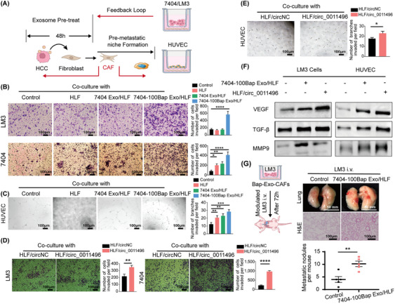
Method: Exosomes were isolated from B[a]P stimulated BEL7404 HCC cells (7404-100Bap Exo) at an environmental relevant dose (100 nmol/L). Lung pre-education animal model was prepared via injection of exosomes and cytokines. The inflammatory genes of educated lungs were evaluated using quantitative reverse transcription PCR array. HCC LM3 cells transfected with firefly luciferase were next injected to monitor tumor burdens and organotropic metastasis. Profile of B[a]P-exposed exosomes were determined by ceRNA microarray. Interactions between circular RNA (circRNA) and microRNAs (miRNAs) were detected using RNA pull-down in target lung fibroblasts. Fluorescence in situ hybridization and RNA immunoprecipitation assay was used to evaluate the "on-off" interaction of circRNA-miRNA pairs. We further developed an adeno-associated virus inhalation model to examine mRNA expression specific in lung, thereby exploring the mRNA targets of B[a]P induced circRNA-miRNA cascade.

Results: Lung fibroblasts exert activation phenotypes, including focal adhesion and motility were altered by 7404-100Bap Exo. In the exosome-educated in vivo model, fibrosis factors and pro-inflammatory molecules of are up-regulated when injected with exosomes. Compared to non-exposed 7404 cells, circ_0011496 was up-regulated following B[a]P treatment and was mainly packaged into 7404-100Bap Exo. Exosomal circ_0011496 were delivered and competitively bound to miR-486-5p in recipient fibroblasts. The down-regulation of miR-486-5p converted fibroblast to cancer-associated fibroblast via regulating the downstream of Twinfilin-1 (TWF1) and matrix metalloproteinase-9 (MMP9) cascade. Additionally, increased TWF1, specifically in exosomal circ_0011496 educated lungs, could promote cancer-stroma crosstalk via activating vascular endothelial growth factor (VEGF). These modulated fibroblasts promoted endothelial cells angiogenesis and recruited primary HCC cells invasion, as a consequence of a pre-metastatic niche formation.

Conclusion: We demonstrated that B[a]P-induced tumor exosomes can deliver circ_0011496 to activate miR-486-5p/TWF1/MMP9 cascade in the lung fibroblasts, generating a feedback loop that promoted HCC metastasis.

Keywords: benzo(a)pyrene; cancer associate fibroblast; circular RNA; exosome; organotropic metastasis.


Conflict of interest statement

The authors declare that they have no competing interests.

Figures

FIGURE 1
FIGURE 1
B[a]P induced HCC‐derived exosomes activate lung fibroblasts. (A) Workflow diagram of the procedure for exosomes extraction and pellet‐free supernatant collection. The exosomes were further co‐culture with fibroblasts for 48 h. (B) Exosomes released by B[a]P treated (7404‐100Bap Exo) and control cells (7404 Exo) were detected by TEM. Scale bar, 100 nm. (C) The size range of exosomes from 7404‐100Bap and 7404 were checked by nanosight particle tracking analysis. (D) Western blotting assay of indicated proteins in exosomes and donor cells. (E) Confocal imaging showed the delivery of SP‐DioC18‐labeled exosomes (green) to Dil‐labeled fibroblasts (red), scale bar, 20μm. (F‐G) qRT‐PCR (F) and Western blotting (G) for the indicated proteins in the HLF cells pre‐treated with 7404 Exo and 7404‐100Bap Exo. (H) Migration transwell assays for HLF treated with equal quantities of exosomes (7404‐100Bap Exo and 7404 Exo) or blank control (PBS). Scale bar, 100 μm. (I) Phalloidin staining of actins in HLF cells pre‐treated with exosomes (7404 Exo and 7404‐100 Bap Exo). Scale bar, 20 μm. **P < 0.01; ***P < 0.001, ****P < 0.0001. Data in F and H were analyzed using one‐way ANOVA with Dunnett's multiple comparisons test. Abbreviations: B[a]P, Benzo(a)pyrene; Con, control; Exo, exosome; HCC, hepatocellular carcinoma; HLF, human derived lung fibroblast; qRT‐PCR,quantitative reverse transcription‐PCR; TEM, transmission electron microscope.
FIGURE 2
FIGURE 2
The contribution of 7404‐100Bap Exo in lung metastasis in vivo. (A) Workflow of lung education by tumor derived exosome in vivo. 7404 Exo and 7404‐100Bap Exo were i.v. injected into mice (n = 8/group) every 3 days for 5 times, the educated lungs were evaluated by PCR array before tumor cells injection. Luciferase‐labeled LM3 cells were next i.v. injected to these educated mice, and the growth and spread of tumor cells were monitored every 7 days till 35 days. (B) Heat map of PCR array for genes encoding cytokines and chemokines were analyzed in the exosome‐educated lung tissues (n = 3/group) after 5 times injection of PBS, 7404 Exo and 7404‐100Bap Exo. Expression values were normalized over the expression of GAPDH and presented as log2 of relative changes. As shown, genes with higher expression are depicted in red, genes with lower expression are depicted in blue. The cytokines and chemokines molecules were grouped according to the pathway in which they participate. (C) The Top 15 dysregulated gene expression network in the pre‐educated lung tissues was analyzed and visualized by String and Cytoscape3.6.1 software. (D) Mice were i.v. injected with 5 × 10⁵ LM3 cells that with stably expressing firefly luciferase (n = 5/group). Representative images and quantitative analysis of lung metastasis of mice using luciferase‐based bioluminescence imaging. (E) The mice in D were sacrificed after 35 days of LM3 injection. H&E and Masson's trichrome staining were detected in the lung tissues harvested from the mice and the IHC results showed the expression levels of α‐SMA. Scale bar, 100μm. (F) Lung metastasis nidus of tumor‐bearing mice after injection of PBS, 7404 Exo and 7404‐100Bap Exo were counted after fixed and stained with bouin's solution. *P < 0.05, ****P < 0.0001. Data in F were analyzed using one‐way ANOVA with Dunnett's multiple comparisons test. Abbreviations: αSMA, alpha smooth muscle actin; GAPDH, Glyceraldehyde‐3‐Phosphate Dehydrogenase; IHC, immunohistochemistry; HCC, hepatocellular carcinoma; H&E, Hematoxylin and eosin; i.v. intravenous.
FIGURE 3
FIGURE 3
Identification of exosomal circ_0011496. (A) ceRNA microarray analysis of the indicated circRNAs in 7404 Exo and 7404‐100Bap Exo. Red and blue represent upregulated and downregulated circRNAs, respectively. (B) Flowchart of cicrRNA‐miRNA‐mRNA network analysis. B[a]P stimulated circRNAs were selected and their sponge miRNAs were predicted 2 databases (circInteractome and circBase). These prediction miRNAs were second screened in the 7404‐100Bap Exo, the down‐regulated miRNAs were selected according to “on‐off” characteristics of circRNA‐miRNA pairs. Interaction of miRNAs and mRNAs were predicted from the intersection of 4 databases (miRwalk, miRDB, TargetScan and miRTarbase). (C) qRT‐PCR analysis of the indicated circRNAs in the control HLF and the HLF pre‐treated with 7404 Exo or 7404‐100Bap Exo. (D) Migration transwell assay of the HLF transfected with the indicated circRNAs. Scale bar, 100 μm. (E) Phalloidin staining showed the adherent morphology of the control HLF and the HLF transfected with circ_0011496. Scale bar, 20 μm. (F) qRT‐PCR analysis of CAF activation‐related genes in the HLF transfected with circ_0011496 cells. (G) The expressions of circ_0011496 in the nuclear, cytoplasmic and cultured medium of 7404‐100Bap cells were detected by qRT‐PCR analysis. ***P < 0.001, ****P < 0.0001. Data in C, D, and G were analyzed using one‐way ANOVA tests with Dunnett's multiple comparisons test. Data in F were analyzed using Student's t‐tests. Abbreviations: BMP, bone morphogenetic protein; CAF, cancer‐associated fibroblast; CCL, chemokine C‐C motif ligand; ceRNA, competing endogenous RNAs; circRNA, circularRNA; HLF, human derived lung fibroblast; IL6, interleukin 6; qRT‐PCR, quantitative reverse transcription PCR; TNF, tumor necrosis factor; VEGF, vascular endothelial growth factor.
FIGURE 4
FIGURE 4
The circular characteristic of circ_0011496 in the CAF activation. (A) Sketch map and Sanger sequencing to confirm the specific back splicing site of circ_0011496 (circCSMD2). (B) qRT‐PCR analysis of circ_0011496 and GAPDH mRNA after RNase R treatment. (C) qRT‐PCR analysis of circ_0011496 and linear CSMD2 mRNA in the tumor cells treated with actinomycin D. (D) The cytoplasmic distribution and location of circ_0011496 in the 7404‐100Bap Exo pre‐treated HLF cells via FISH assay. Scale bar, 20μm. (E) 7404‐100Bap cells were transfected with siRNA‐control (siNC) and 2 circ_0011496 siRNAs (si‐ circ_0011496‐01 and si‐ circ_0011496‐02). Silencing efficiency of two circ_0011496 siRNAs in these 7404‐100Bap cells were detected by qRT‐PCR. (F) Exosomes isolated from 7404‐100Bap cells that transfected with siNC or circ_0011496 siRNAs (7404‐100Bap siNC/Exo and 7404‐100Bap si‐circ_0011496‐01/02/Exo), and expressions of exosomal circ_0011496 were evaluated by qRT‐PCR. (G) Migration transwell assay of HLF cells treated with the exosomes derived from 7404‐100Bap cells (Bap Exo‐siNC) and 7404‐100Bap/si‐circ_0011496‐02 cells (Bap Exo‐si‐ circ_0011496). (H) Exosomes, Bap Exo‐siNC and Bap Exo‐si‐circ_0011496, were collected (200μg/mL) and then i.v. injected to mice every 3days for 5 times to generate circ_0011496‐deficient lungs (n = 8/group). LM3 cells (5 × 10⁵) were further i.v. injected and the tumor metastasis were monitored till 28 days. (I) Representative FISH and IHC images for circ_0011496 and fibroblast markers (vimentin) in the same areas of these lungs after 14 days of Bap Exo‐siNC and Bap Exo‐si‐circ_0011496 education. Scale bar, 20 μm. (J) Luciferase‐based bioluminescence imaging of metastatic LM3 cells after 28 days i.v. injection in the exosome‐educated models to determine the key role of circ_0011496 in promoting lung metastasis. *P < 0.05; **P < 0.01, ***P < 0.001. Data in E and F were analyzed using one‐way ANOVA tests with Dunnett's multiple comparisons test. Data in B, C, and G were analyzed using Student's t‐tests. Abbreviations: CSMD2, CUB and Sushi multiple domains 2; FISH, fluorescence in situ hybridization; IHC, immunohistochemistry; i.v., intravenous injection; qRT‐PCR, quantitative reverse transcription‐PCR; siRNA, small interfering RNA.
FIGURE 5
FIGURE 5
circ_0011496 served as a molecular sponge for miR‐486‐5p. (A) Volcano plot illustrates the profile of sponges’ miRNAs obtained by Biotin‐labeled RNA pull‐down assay of circ_0011496 probe in the 7404‐100Bap Exo educated‐HLF cells comparing with HLF cells. |log2(Fold change)| ≥1 and P < 0.05). (B)The sponges’ miRNAs were screened by pull down of circ_0011496 specific probe in the total RNA input of HLF cells (HLF‐IP) and the small RNA of HLF cells (HLF‐P). The top 10 expression miRNAs in the intersection (HLF‐IP & HLF‐P) that were identified as specifically sponges bind to circ_0011496. (C) Northern blotting performed to detect interaction between circ_0011496 and miR‐486‐5p. miRNA‐486‐5p was pulled down by the circ_0011496 probe (circ‐probe) but not its mutant random probe (ran‐probe). (D) 7404‐100Bap cells were transfected with si‐NC, si‐ circ_0011496 and the overexpression plasmid of circ_0011496 (Ov‐circ_0011496), and their derived exosomes were further collected, respectively. HLF cells were co‐cultured with these different exosomes (HLF + Exo‐si‐circ_0011496, HLF + Exo‐ov‐circ_0011496,) for 48h, and the expression levels of mir‐486‐5p were evaluated by qRT‐PCR assay. (E) A schematic of wild‐type (WT) and mutant (MUT) circ_0011496 luciferase reporter vectors. Dual‐luciferase reporter assay was performed to assess the interaction between circ_0011496 WT or circ_0011496 MUT of circ_0011496 and miR‐486‐5p. (F) Anti‐AGO2 RIP assays were used in 7404‐100Bap Exo modulated fibroblasts to determine the enrichment of circ_0011496 and miR‐486‐5p. Anti‐IgG was used as control. (G) qRT‐PCR analysis of the expression levels of miR‐486‐5p in the lung fibroblasts treated with7404‐100Bap Exo at indicated time points. (H) HLF control, HLF treated with 7404‐100Bap Exo (HLF + 7404‐100Bap Exo), HLF transfected with circ_0011496 (HLF + circ_0011496), and HLF treated with exosomes derived form 7404‐100Bap cells‐transfected si‐circ_0011496 (HLF + BapExo‐siCirc) were used as cell models. FISH indicates the colocation of circ_0011496 and miR‐486‐5p in these cell models, circ_0011496 probe was labeled red with biotin. Scale bar, 20 μm. *P < 0.05; ***P < 0.001; ns, not significant. Data in F and G were analyzed using one‐way ANOVA tests with Dunnett's multiple comparisons test. Data in D and E were analyzed using Student's t‐tests. Abbreviations: FISH, fluorescence in situ hybridization; HLF, human derived lung fibroblast; IHC, immunohistochemistry; i.v., intravenous injection; qRT‐PCR, quantitative reverse transcription‐PCR; RIP, RNA immunoprecipitation; siRNA, small interfering RNA.
FIGURE 6
FIGURE 6
TWF1 is a direct downstream target of miR‐486‐5p. (A) Target gene prediction of miR‐486‐5p with four bioinformatics tools. (B) TWF1 expressions were evaluated in the control HLF cells and 7404‐100Bap Exo pre‐treated HLF cells (HLF + Exo). For in vivo assessment, i. v. injection of PBS control (Lung) and 7404‐100Bap Exo (Lung + Exo) to mice for 15 days to generated an education model as described, and TWF1 expressions of these lungs were evaluated by qRT‐PCR. (C) qRT‐PCR analysis of TWF1 expression in the HLF cells transfected with miR‐486‐5p mimic or control miRNA (miR‐NC), respectively. (D) Relative luciferase activity of the wild‐type (WT) and mutant type (MUT) of miR‐486‐5p binding site in TWF1 3’UTR. (E) Western blotting of indicated proteins in 7404 Exo or 7404‐100Bap Exo treated HLF cells. (F) Western‐blotting of indicated proteins in HLF cells transfected with miR‐486‐5p mimic, circ_0011496 overexpression plasmid, and TWF1 siRNA (siTWF1). (G) Migration transwell assay for HLF transfected with siTWF1 and miR‐486‐5p mimic. Scale bar, 100 μm. (H) Schematic illustration for the in vivo delivery of AAV‐mediated TWF1 overexpression in lung and lung metastasis analysis. TWF1 transfected into the AAV vectors (50μg/mL) and introduced to mice via inhalation every 5‐6 days for 5 times to specific express TWF1 in lungs. LM3 cells (5 × 10⁵) were i.v. injected by the 14 days of TWF1‐AAV inhalation, and the tumor growth and metastasis were analyzed at the end of 40 days. (I‐J) Mice along with lung metastasis mention in H were sacrificed 40 days after LM3 injection. H&E staining of lung tissues (I) and the images of HCC tumor‐bearing lung (J) were shown. *P < 0.05; **P < 0.01, ***P < 0.001, ****P < 0.0001. Scale bar, 100 μm.Data in G were analyzed using one‐way ANOVA tests with Dunnett's multiple comparisons test. Data in B, C, and D were analyzed using Student's t‐tests. Abbreviations: 3’UTR, 3' untranslated regions; AAV, adeno‐associated virus; HLF, human derived lung fibroblast; IL11, interleukin; i.v., intravenous; MMP9, matrix metalloproteinase 9; STAT3, signal transducer and activator of transcription 3; TWF1, twinfilin‐1.
FIGURE 7
FIGURE 7
CAFs promote angiogenesis of HUVEC and migration of LM3 exoaomal circRNA. (A) Schematic diagram of Exo‐modulated fibroblast cells regulated surrounding stroma cells, and generated a feedback loop for distant tumor cells. The HLF cells were pre‐treated with 7404 Exo or 7404‐100Bap Exo for 48h for activation (7404Exo/HLF, 7404‐100Bap Exo/HLF), these activated‐HLF cells were next co‐cultured with 7404 and LM3 cells for transwell assay, and with HUVEC cells for angiogenesis tube formation. (B) Migration of HCC 7404 and LM3 cells co‐cultured with PBS control, HLF cells, 7404Exo/HLF cells, 7404‐100Bap Exo/HLF cells were accessed by transwell assay. Scale bar, 100 μm. (C) Angiogenesis ability of HUVEC cells co‐cultured with PBS control, HLF cells, 7404Exo/HLF cells, 7404‐100Bap Exo/HLF cells were accessed by matrigel‐based tube formation. Scale bar, 100 μm. (D) Migration of HCC cells treated with HLF‐transfected circ_0011496 vector was accessed by transwell assay. Scale bar, 100 μm. (E) Tube formation ability of HUVEC treated with HLF‐transfected circ_0011496 vector. Scale bar, 100 μm. (F) Western blotting of indicated proteins in the HLF cells co‐cultured 7404‐100Bap Exo, as well as co‐cultured with HLF‐transfected circ_0011496 vector. (G) We developed an in vivo experiment to explore the cancer‐stroma crosstalk, LM3 cells and 7404‐100Bap Exo/HLF cells were co‐cultured using 0.4μm‐transwell chamber for 72h, so that LM3 cells could not pass through the pore but effected by signal transduction. These co‐cultured LM3 cells were i.v. injected into mice, and the lungs after 35 days LM3 injection were scarfed and evaluated by H&E staining. Scale bar, 100 μm for H&E and 50mm for lung. *P < 0.05; **P < 0.01, ***P < 0.001, ****P < 0.0001. Data in B and C were analyzed using one‐way ANOVA tests with Dunnett's multiple comparisons test. Data in E and D were analyzed using Student's t‐tests. Abbreviations: CAF, cancer‐associated fibroblast; HCC, hepatocellular carcinoma; H&E, hematoxylin and eosin; HUVEC, human umbilical vein endothelial cell; MMP9, matrix metalloproteinases 9; STAT3, signal transducer and activator of transcription 3; TGF‐β, transforming growth factor‐β.
FIGURE 8
FIGURE 8
Schematic representation of the role of B[a]P in HCC lung‐specific metastasis. The schematic shows the effect of B[a]P in HCC lung‐specific metastasis through exosomal circRNA. Before tumor cell arriving, exposure of B[a]P trigger an increased in the amount of exosomal circRNA to target lung fibroblasts, contributing to the transformation of fibroblasts in the metastatic organ. These Exo‐activated fibroblasts further generate a feedback loop either for epithelial cell angiogenesis or recruitment of primary HCC invasion. Abbreviations: B[a]P, Benzo[a]pyrene; HCC, hepatocellular carcinoma.

References

    1. Lange NF, Radu P, Dufour JF. Prevention of NAFLD‐associated HCC: role of lifestyle and chemoprevention. J Hepatol. 2021(5):1217–1227. - PubMed
    1. Huang DQ, El‐Serag HB, Loomba R. Global epidemiology of NAFLD‐related HCC: trends, predictions, risk factors and prevention. Nat Rev Gastroenterol Hepatol. 2020;18(4):223–238. - PMC - PubMed
    1. Ma Y, Yang W, Li T, Liu Y, Simon TG, Sui J, et al. Meat intake and risk of hepatocellular carcinoma in two large US prospective cohorts of women and men. Int J Epidemiol. 2019;48(6):1863–1871. - PMC - PubMed
    1. Wang Z, Yang P, Xie J, Lin HP, Yang C. Arsenic and benzo[a]pyrene co‐exposure acts synergistically in inducing cancer stem cell‐like property and tumorigenesis by epigenetically down‐regulating SOCS3 expression. Environ Int. 2020;137:105560. - PMC - PubMed
    1. Wu HC, Wang Q, Wang LW, Yang HI, Ahsan H, Tsai WY, et al. Polycyclic aromatic hydrocarbon‐ and aflatoxin‐albumin adducts, hepatitis B virus infection and hepatocellular carcinoma in Taiwan. Cancer Lett. 2007;252(1):104–114. - PMC - PubMed

MeSH terms