Spinal epidermoid cyst associated with limited dorsal myeloschisis
- PMID: 38840610
- PMCID: PMC11152525
- DOI: 10.25259/SNI_291_2024
Spinal epidermoid cyst associated with limited dorsal myeloschisis
Abstract
Background: Epidermoid cysts (ECs) are rare benign tumors arising from epidermal cells, associated with congenital abnormalities or acquired through trauma, surgery, or lumbar punctures. They represent <1% of all intraspinal tumors and may be associated with limited dorsal myeloschisis (LDM).
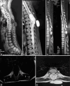
Case description: A 7-year-old neurologically intact male had a dorsal skin mass since birth located posteriorly in the midline of the inferior thoracic spine. The mass was palpable, painless, mobile, vascularized, and could be transilluminated. Thoracic magnetic resonance imaging showed an extensive intradural extramedullary cystic lesion extending from D6 to D8 that did not enhance with contrast, accompanied by a subcutaneous fluid collection at D8-D9 communicating with the subarachnoid space. The patient underwent gross total resection of the lesion, pathologically confirmed as an EC. The postoperative course was uneventful, with no recurrence 1 year postoperatively.
Conclusion: LDM may be associated with ECs. Early diagnosis and surgical resection of these lesions are essential for favorable outcomes.
Keywords: Congenital epidermoid cyst; Limited dorsal myeloschisis; Spine.
Copyright: © 2024 Surgical Neurology International.
Conflict of interest statement
There are no conflicts of interest.
Figures


Similar articles
-
Dorsal Spinal Intradural Intramedullary Epidermoid Cyst: A Rare Case Report and Review of Literature.J Neurosci Rural Pract. 2019 Apr-Jun;10(2):352-354. doi: 10.4103/jnrp.jnrp_304_18. J Neurosci Rural Pract. 2019. PMID: 31001035 Free PMC article.
-
Thoracic Intradural-Extramedullary Epidermoid Tumor: The Relevance for Resection of Classic Subarachnoid Space Microsurgical Anatomy in Modern Spinal Surgery. Technical Note and Review of the Literature.World Neurosurg. 2017 Dec;108:54-61. doi: 10.1016/j.wneu.2017.08.078. Epub 2017 Aug 24. World Neurosurg. 2017. PMID: 28843754 Review.
-
Acquired dorsal intraspinal epidermoid cyst in an adult female.Surg Neurol Int. 2016 Jan 25;7(Suppl 3):S67-9. doi: 10.4103/2152-7806.174890. eCollection 2016. Surg Neurol Int. 2016. PMID: 26904369 Free PMC article.
-
Limited Dorsal Myeloschisis and Congenital Dermal Sinus: Comparison of Clinical and MR Imaging Features.AJNR Am J Neuroradiol. 2017 Jan;38(1):176-182. doi: 10.3174/ajnr.A4958. Epub 2016 Oct 20. AJNR Am J Neuroradiol. 2017. PMID: 27765739 Free PMC article.
-
A rare case of intradural and extramedullary epidermoid cyst after repetitive epidural anesthesia: case report and review of the literature.World J Surg Oncol. 2017 Jul 17;15(1):131. doi: 10.1186/s12957-017-1186-4. World J Surg Oncol. 2017. PMID: 28716031 Free PMC article. Review.
Cited by
-
Spinal Cord Müllerian-Like Cyst at the T12 Vertebral Level: A Case Report and Literature Review.Cureus. 2025 Mar 6;17(3):e80169. doi: 10.7759/cureus.80169. eCollection 2025 Mar. Cureus. 2025. PMID: 40190987 Free PMC article.
References
-
- Bansal S, Suri A, Borkar SA, Kale SS, Singh M, Mahapatra AK. Management of intramedullary tumors in children: Analysis of 82 operated cases. Childs Nerv Syst. 2012;28:2063–9. - PubMed
-
- Bretz A, Van den Berge D, Storme G. Intraspinal epidermoid cyst successfully treated with radiotherapy: Case report. Neurosurgery. 2003;53:1429–31. discussion 1431-2. - PubMed
-
- Dobre MC, Smoker WR, Moritani T, Kirby P. Spontaneously ruptured intraspinal epidermoid cyst causing chemical meningitis. J Clin Neurosci. 2012;19:587–9. - PubMed
-
- Erşahin Y, Barçin E, Mutluer S. Is meningocele really an isolated lesion? Childs Nerv Syst. 2001;17:487–90. - PubMed
-
- Graillon T, Rakotozanany P, Meyer M, Dufour H, Fuentes S. Intramedullary epidermoid cysts in adults: Case report and updated literature review. Neurochirurgie. 2017;63:99–102. - PubMed
Publication types
LinkOut - more resources
Full Text Sources
