

Preoperative Degenerative Changes at the Tibial Sesamoid-Metatarsal Joint in Hallux Valgus: Association With Postoperative Patient-Reported Outcomes After Modified Lapidus Procedure
- PMID: 38840786
- PMCID: PMC11151770
- DOI: 10.1177/24730114241256370
Preoperative Degenerative Changes at the Tibial Sesamoid-Metatarsal Joint in Hallux Valgus: Association With Postoperative Patient-Reported Outcomes After Modified Lapidus Procedure
Abstract
Background: Degenerative changes at the sesamoid-metatarsal joints (SMJs) may be a source of pain following hallux valgus surgery. The aims of this study were to describe degenerative changes at the SMJs on weightbearing computed tomography (WBCT) scans and, secondarily, investigate their association with 1-year patient-reported outcome scores following a modified Lapidus procedure for hallux valgus. We hypothesized that reduced joint space in the SMJs would correlate with worse patient-reported outcomes.
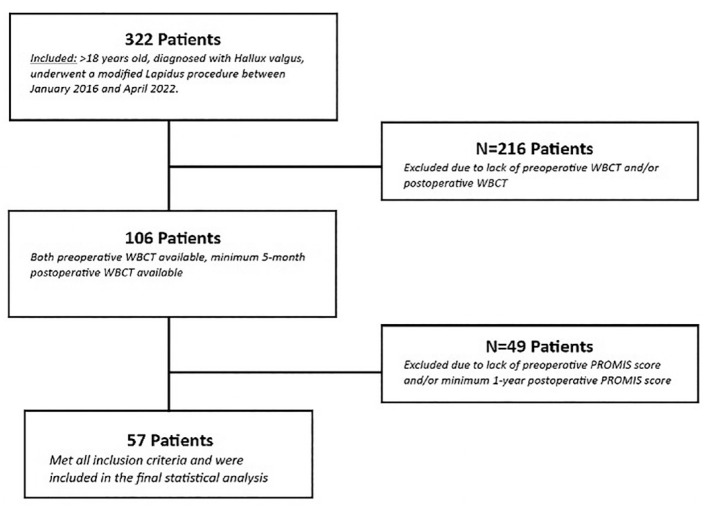
Methods: Fifty-seven hallux valgus patients who underwent a modified Lapidus procedure had preoperative and minimum 5-month postoperative WBCT scans, and preoperative and at least 1-year postoperative PROMIS physical function (PF), pain interference, and pain intensity scores were included. Degenerative changes at the SMJs were measured using distance mapping between the sesamoids and first metatarsal head on preoperative and postoperative WBCT scans. The minimum and average distances between the first metatarsal head and tibial sesamoid (tibial-SMJ) for each patient preoperatively and postoperatively were measured. Sesamoid station was measured on WBCT scans using a 0 to 3 grading system. Linear regression was used to investigate the correlations between minimum preoperative and postoperative tibial-SMJ distances and 1-year postoperative PROMIS scores.
Results: The median minimum and average tibial-SMJ distances increased from 0.82 mm (interquartile range [IQR] 0.40-1.03 mm) and 1.62 mm (IQR 1.37-1.75 mm) preoperative to 1.09 mm (IQR 0.96-1.23 mm) and 1.73 mm (IQR 1.60-1.91 mm) postoperative (P < .001 and P < .001), respectively. In a subset of patients with complete sesamoid reduction, we found an association between preoperative minimum tibial-SMJ distance and 1-year postoperative PROMIS PF scores (coefficient 7.2, P = .02).
Conclusion: Following the modified Lapidus procedure, there was a statistically significant increase in the tibial-SMJ distance. Additionally, in patients with reduced sesamoids postoperatively, reduced preoperative tibial-SMJ distance correlated with worse PROMIS PF scores.
Level of evidence: Level IV, case series.
Keywords: hallux valgus; metatarsosesamoid joint; modified Lapidus procedure; patient-reported outcomes.
© The Author(s) 2024.
Conflict of interest statement
The author(s) declared no potential conflicts of interest with respect to the research, authorship, and/or publication of this article. Disclosure forms for all authors are available online.
Figures


Similar articles
-
Relationship Between Displacement and Degenerative Changes of the Sesamoids in Hallux Valgus.Foot Ankle Int. 2016 Dec;37(12):1303-1309. doi: 10.1177/1071100716661827. Epub 2016 Aug 16. Foot Ankle Int. 2016. PMID: 27530982
-
Correlation of Clinical Outcomes and Relative Position of the First Metatarsal After the Modified Lapidus Procedure.Foot Ankle Int. 2024 Sep;45(9):979-987. doi: 10.1177/10711007241255378. Epub 2024 Jun 13. Foot Ankle Int. 2024. PMID: 38872316
-
Association Between Postoperative Medial-Middle Intercuneiform Joint Widening and Recurrence Rates in Hallux Valgus Treated With Modified Lapidus Procedure.Foot Ankle Int. 2024 Dec;45(12):1349-1358. doi: 10.1177/10711007241286890. Epub 2024 Nov 11. Foot Ankle Int. 2024. PMID: 39526789
-
Effect of the Modified Lapidus Procedure on Pronation of the First Ray in Hallux Valgus.Foot Ankle Int. 2020 Feb;41(2):125-132. doi: 10.1177/1071100719883325. Epub 2019 Oct 16. Foot Ankle Int. 2020. PMID: 31617413
-
Relationship of frontal plane rotation of first metatarsal to proximal articular set angle and hallux alignment in patients undergoing tarsometatarsal arthrodesis for hallux abducto valgus: a case series and critical review of the literature.J Foot Ankle Surg. 2013 May-Jun;52(3):348-54. doi: 10.1053/j.jfas.2013.01.006. Epub 2013 Mar 6. J Foot Ankle Surg. 2013. PMID: 23473673 Review.
References
-
- Athanasiou KA, Liu GT, Lavery LA, Lanctot DR, Schenck RC., Jr. Biomechanical topography of human articular cartilage in the first metatarsophalangeal joint. Clin Orthop Relat Res. 1998;348:269-281. - PubMed
-
- Bock P, Kluger R, Kristen KH, Mittlböck M, Schuh R, Trnka HJ. The scarf osteotomy with minimally invasive lateral release for treatment of hallux valgus deformity: intermediate and long-term results. J Bone Joint Surg Am. 2015;97(15):1238-1245. - PubMed
-
- Campbell B, Miller MC, Williams L, Conti SF. Pilot study of a 3-dimensional method for analysis of pronation of the first metatarsal of hallux valgus patients. Foot Ankle Int. 2018;39(12):1449-1456. - PubMed
-
- Chen JY, Rikhraj K, Gatot C, Lee JY, Singh Rikhraj I. Tibial sesamoid position influence on functional outcome and satisfaction after hallux valgus surgery. Foot Ankle Int. 2016;37(11):1178-1182. - PubMed
LinkOut - more resources
Full Text Sources
Research Materials