. 2024 May 22:18:1340468.
doi: 10.3389/fnana.2024.1340468. eCollection 2024.

Division of neuromuscular compartments and localization of the center of the highest region of muscle spindles abundance in deep cervical muscles based on Sihler's staining

Affiliations

Division of neuromuscular compartments and localization of the center of the highest region of muscle spindles abundance in deep cervical muscles based on Sihler's staining

Danli Wang et al. Front Neuroanat. .

Abstract

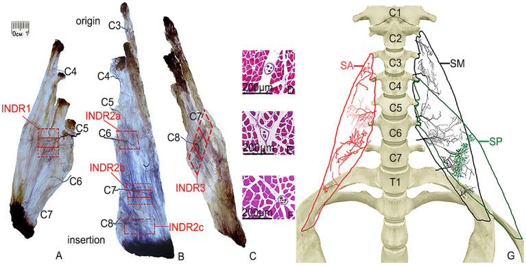
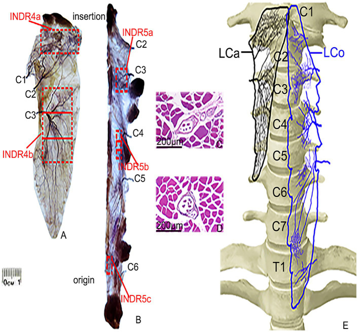
Purpose: The overall distribution pattern of intramuscular nerves and the regions with the highest spindle abundance in deep cervical muscles have not been revealed. This study aimed to reveal neuromuscular compartmentalization and localize the body surface position and depth of the center of the region of highest muscle spindle abundance (CRHMSA) in the deep cervical muscles.

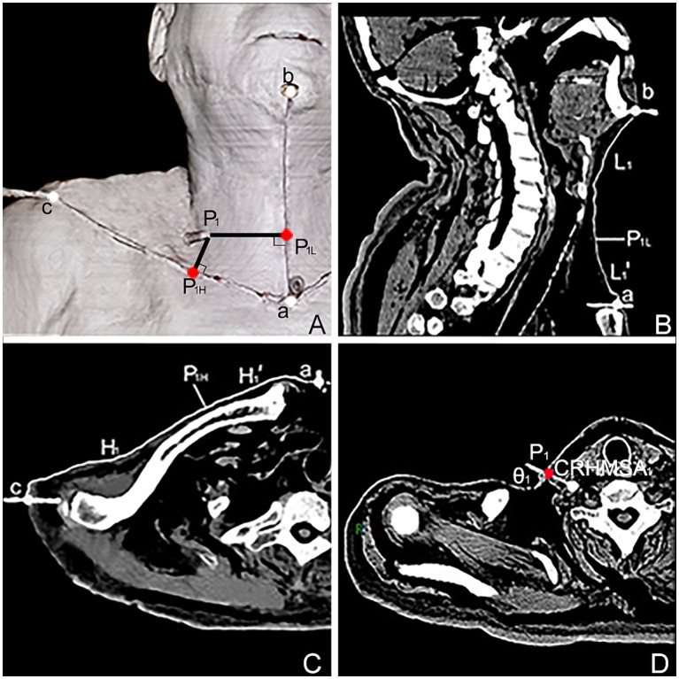
Methods: This study included 36 adult cadavers (57.7 ± 11.5 years). The curved line joining the lowest point of the jugular notch and chin tip was designated as the longitudinal reference line (line L), and the curved line connecting the lowest point of the jugular notch and acromion was designated as the horizontal reference line (line H). Modified Sihler's staining, hematoxylin-eosin staining and computed tomography scanning were employed to determine the projection points (P) of the CRHMSAs on the anterior surfaces of the neck. The positions (PH and PL) of point P projected onto the H and L lines, and the depth of each CRHMSA, and puncture angle were determined using the Syngo system.

Results: The scalenus posterior and longus capitis muscles were divided into two neuromuscular compartments, while the scalenus anterior and longus colli muscles were divided into three neuromuscular compartments. The scalenus medius muscle can be divided into five neuromuscular compartments. The PH of the CRHMSA of the scalenus muscles (anterior, medius, and posterior), and longus capitis and longus colli muscles, were located at 36.27, 39.18, 47.31, 35.67, and 42.71% of the H line, respectively. The PL positions were at 26.53, 32.65, 32.73, 68.32, and 51.15% of the L line, respectively. The depths of the CRHMSAs were 2.47 cm, 2.96 cm, 2.99 cm, 3.93 cm, and 3.17 cm, respectively, and the puncture angles were 87.13°, 85.92°, 88.21°, 58.08°, and 77.75°, respectively.

Conclusion: Present research suggests that the deep cervical muscles can be divided into neuromuscular compartments; we recommend the locations of these CRHMSA as the optimal target for administering botulinum toxin A injections to treat deep cervical muscle dystonia.

Keywords: Sihler’s staining; deep cervical muscle; muscle spindle; neuromuscular compartment; target localization.


Conflict of interest statement

The authors declare that the research was conducted in the absence of any commercial or financial relationships that could be construed as a potential conflict of interest. The reviewer PX declared a shared affiliation with the authors to the handling editor at the time of review.

Figures

Figure 1
Figure 1
Sihler’s staining of the SA, SM, and SP muscles and representative muscle spindles in the highest region of muscle spindle abundance. (A–C) Sihler staining of the SA, SM, and SP muscles (superficial view). Red boxes indicate INDR1, INDR2a, INDR2b, INDR2c, and INDR3. (D–F) Representative muscle spindles with the highest abundance of INDR1, INDR2b, and INDR3 (scale: 200 μm). (G) Distribution pattern of the intramuscular nerves of the SA, SM, SP are shown in red, black, and green, respectively.
Figure 2
Figure 2
Sihler’s staining and representative muscle spindles in the highest region of muscle spindle abundance in the LCa and LCo muscles. (A,B) Sihler’s staining of the LCa and LCo (superficial view). Red boxes indicate INDR4a, INDR4b, INDR5a, INDR5b, and INDR5c. (C,D) Representative muscle spindles with the highest abundance in INDR4b and INDR5b (scale: 200 μm). (E) Distribution pattern of the intramuscular nerves of the LCa and LCo are shown in black and blue, respectively.
Figure 3
Figure 3
Simulated puncture of right SA muscle CRHMSA. 1: SA muscle, 2: phrenic nerve, 3: ascending cervical artery, 4: 5th cervical nerve (C5), 5: transverse cervical artery, 6: SM muscle, 7: SP muscle, 8: needle.
Figure 4
Figure 4
Spiral CT localization image of the center of the highest region of muscle spindle abundance (CRHMSA1) of the right SA muscle in males. (A) Three-dimensional reconstructed spiral CT image showing the position of CRHMSA1 on the body surface and the designed reference line. P1 is the surface projection point of the SA muscle CRHMSA1. P1H = intersection point generated by creating a line perpendicular to line H through P1. P1L = intersection point of the H-line passing through P1 and the L-line. a-P1H=H1’, a-P1L = L1’. (B) Lengths of the L and L1 lines in the coronal plane were measured. (C) Measure the lengths of the H and H1’ lines on the section passing through line H. (D) Determine the depth and angle of needle insertion for CRHMSA1 in the cross section.

Similar articles

Cited by

References

    1. Allison S. K., Odderson I. R. (2016). Ultrasound and electromyography guidance for injection of the longus Colli with botulinum toxin for the treatment of cervical dystonia. Ultrasound Q. 32, 302–306. doi: 10.1097/RUQ.0000000000000226, PMID: - DOI - PubMed
    1. Amirali A., Mu L., Gracies J. M., Simpson D. M. (2007). Anatomical localization of motor endplate bands in the human biceps brachii. J. Clin. Neuromuscul. Dis. 9, 306–312. doi: 10.1097/CND.0b013e31815c13a7, PMID: - DOI - PubMed
    1. Baltopoulos P., Tsintzos C., Prionas G., Tsironi M. (2008). Exercise-induced scalenus syndrome. Am. J. Sports Med. 36, 369–374. doi: 10.1177/0363546507312166, PMID: - DOI - PubMed
    1. Banks R. W. (2006). An allometric analysis of the number of muscle spindles in mammalian skeletal muscles. J. Anat. 208, 753–768. doi: 10.1111/j.1469-7580.2006.00558.x, PMID: - DOI - PMC - PubMed
    1. Borodic G. E., Ferrante R., Pearce L. B., Smith K. (1994). Histologic assessment of dose-related diffusion and muscle fiber response after therapeutic botulinum a toxin injections. Mov. Disord. 9, 31–39. doi: 10.1002/mds.870090106, PMID: - DOI - PubMed

LinkOut - more resources