

Cellular interactions and microenvironment dynamics in skeletal muscle regeneration and disease
- PMID: 38840849
- PMCID: PMC11150574
- DOI: 10.3389/fcell.2024.1385399
Cellular interactions and microenvironment dynamics in skeletal muscle regeneration and disease
Abstract
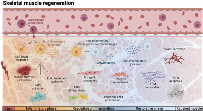
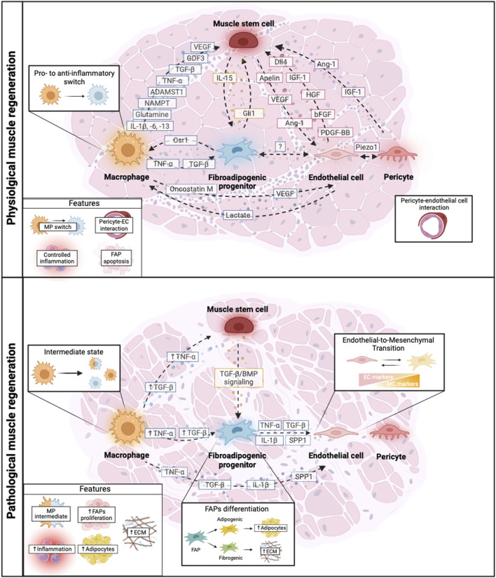
Skeletal muscle regeneration relies on the intricate interplay of various cell populations within the muscle niche-an environment crucial for regulating the behavior of muscle stem cells (MuSCs) and ensuring postnatal tissue maintenance and regeneration. This review delves into the dynamic interactions among key players of this process, including MuSCs, macrophages (MPs), fibro-adipogenic progenitors (FAPs), endothelial cells (ECs), and pericytes (PCs), each assuming pivotal roles in orchestrating homeostasis and regeneration. Dysfunctions in these interactions can lead not only to pathological conditions but also exacerbate muscular dystrophies. The exploration of cellular and molecular crosstalk among these populations in both physiological and dystrophic conditions provides insights into the multifaceted communication networks governing muscle regeneration. Furthermore, this review discusses emerging strategies to modulate the muscle-regenerating niche, presenting a comprehensive overview of current understanding and innovative approaches.
Keywords: endothelial cells; fibro-adipogenic progenitors; macrophages; muscle niche dynamics; muscle stem cells; muscular dystrophies; regenerative medicine strategies; skeletal muscle regeneration.
Copyright © 2024 Rodríguez, Timóteo-Ferreira, Minchiotti, Brunelli and Guardiola.
Conflict of interest statement
The authors declare that the research was conducted in the absence of any commercial or financial relationships that could be construed as a potential conflict of interest.
Figures
Similar articles
-
Signaling pathways regulating the fate of fibro/adipogenic progenitors (FAPs) in skeletal muscle regeneration and disease.FEBS J. 2022 Nov;289(21):6484-6517. doi: 10.1111/febs.16080. Epub 2021 Jul 6. FEBS J. 2022. PMID: 34143565 Review.
-
Fibro-adipogenic progenitors in skeletal muscle homeostasis, regeneration and diseases.Open Biol. 2021 Dec;11(12):210110. doi: 10.1098/rsob.210110. Epub 2021 Dec 8. Open Biol. 2021. PMID: 34875199 Free PMC article. Review.
-
Muscle fibro-adipogenic progenitors from a single-cell perspective: Focus on their "virtual" secretome.Front Cell Dev Biol. 2022 Sep 19;10:952041. doi: 10.3389/fcell.2022.952041. eCollection 2022. Front Cell Dev Biol. 2022. PMID: 36200044 Free PMC article.
-
Odd skipped-related 1 controls the pro-regenerative response of fibro-adipogenic progenitors.NPJ Regen Med. 2023 Apr 5;8(1):19. doi: 10.1038/s41536-023-00291-6. NPJ Regen Med. 2023. PMID: 37019910 Free PMC article.
-
Control of Muscle Fibro-Adipogenic Progenitors by Myogenic Lineage is Altered in Aging and Duchenne Muscular Dystrophy.Cell Physiol Biochem. 2019;53(6):1029-1045. doi: 10.33594/000000196. Cell Physiol Biochem. 2019. PMID: 31865646
Cited by
-
Exosomal miR-24-3p mediates myoblast-macrophage crosstalk to promote abdominal muscle repair.Front Pharmacol. 2025 Jun 3;16:1604776. doi: 10.3389/fphar.2025.1604776. eCollection 2025. Front Pharmacol. 2025. PMID: 40529508 Free PMC article.
-
Engineering of tissue in microphysiological systems demonstrated by modelling skeletal muscle.Regen Biomater. 2025 Jun 16;12:rbaf059. doi: 10.1093/rb/rbaf059. eCollection 2025. Regen Biomater. 2025. PMID: 40717794 Free PMC article. Review.
-
In the Era of Cardiovascular-Kidney-Metabolic Syndrome in Cardio-Oncology: From Pathogenesis to Prevention and Therapy.Cancers (Basel). 2025 Mar 30;17(7):1169. doi: 10.3390/cancers17071169. Cancers (Basel). 2025. PMID: 40227756 Free PMC article. Review.
References
Publication types
LinkOut - more resources
Full Text Sources