

An insight into the epidemiology of foodborne zoonotic fascioliasis in small ruminants in northwestern region of Bangladesh
- PMID: 38840875
- PMCID: PMC11147978
- DOI: 10.1007/s12639-024-01672-4
An insight into the epidemiology of foodborne zoonotic fascioliasis in small ruminants in northwestern region of Bangladesh
Abstract
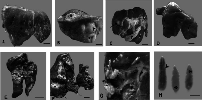
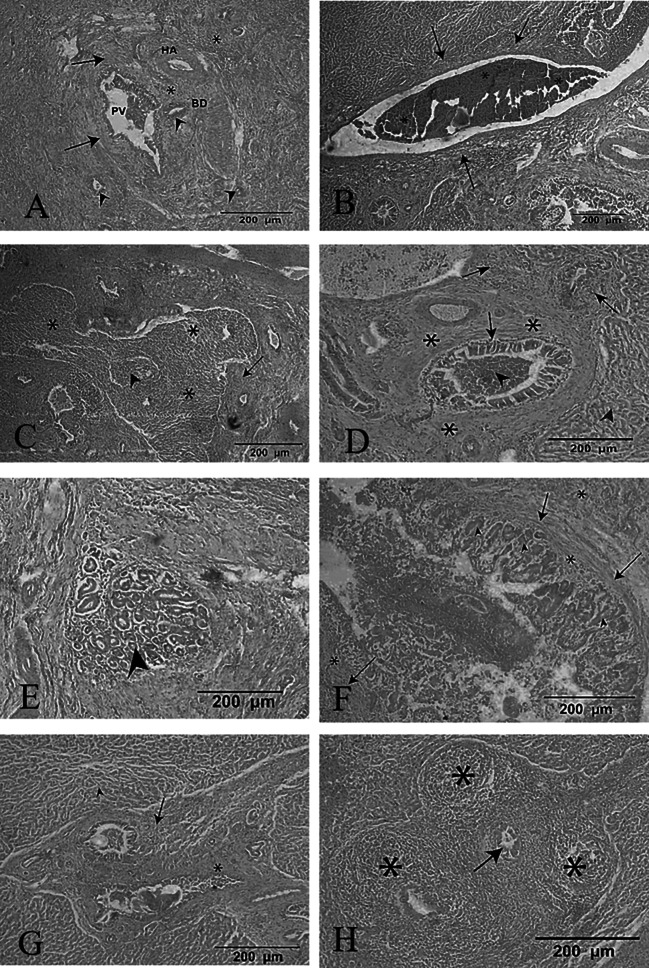
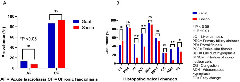
Fascioliasis is one of the most common foodborne zoonotic infection of ruminants in Bangladesh. To estimate the prevalence and associated risk factors of fascioliasis and extent of liver damage, 825 livers of sheep and goats were randomly inspected during onsite slaughterhouse visiting in Naogaon, Natore, Rajshahi and Joypurhat districts. The overall prevalence of fascioliasis was 25.09% and significantly (P = 0.008) higher in goats (26.11%) than sheep (24.00%). During gross inspection, Fasciola infected livers were increased in size, fibrosed, fatty, multiple white or reddish necrotic foci on the parietal surface, hard to cut, calcified, and numerous mature and immature flukes were also observed. In histoarchitecture, inflammatory cell infiltration in the hepatic parenchyma and periportal area, fibrous connective tissue proliferation around necrotic area, hyperplastic bile duct, congestion, and primary biliary cirrhosis were seen in acute and chronic fascioliasis. Epidemiological investigations revealed that fascioliasis was higher in goats than sheep. Age, sex, BCS and season were found to have statistically significant associations with fascioliasis in goats. In case of sheep, age (OR = 5.8671; 95% CI: 2.9482-11.6757, P < 0.0001), sex (OR = 3.7317; 95% CI: 1.9052-7.3094, p < 0.0001), BCS (OR = 6.0346; 95% CI: 1.7986-20.2472, p < .0001), and season (OR = 8.2308; 95% CI: 3.9922-16.9693, p = < .0001) were also found to have statistically significant associations with fascioliasis. Results of the study can help for molecular epidemiology of fascioliasis in small ruminants to plan fluke control programs for safe food production.
Keywords: Bangladesh; Epidemiology; Fascioliasis; Goat; Sheep; Slaughterhouse.
© Indian Society for Parasitology 2024. Springer Nature or its licensor (e.g. a society or other partner) holds exclusive rights to this article under a publishing agreement with the author(s) or other rightsholder(s); author self-archiving of the accepted manuscript version of this article is solely governed by the terms of such publishing agreement and applicable law.
Conflict of interest statement
Conflict of interestThe authors declare that they have no conflict of interests related to this work. They are solely accountable for the content and writing of the report.
Figures
Similar articles
-
Fascioliasis may promote tuberculous infectivity in small ruminants.Saudi J Biol Sci. 2022 Oct;29(10):103402. doi: 10.1016/j.sjbs.2022.103402. Epub 2022 Aug 6. Saudi J Biol Sci. 2022. PMID: 36039324 Free PMC article.
-
Prevalence and hepatic histopathological findings of fascioliasis in sheep slaughtered in Jeddah, Saudi Arabia.Sci Rep. 2023 Apr 24;13(1):6609. doi: 10.1038/s41598-023-33927-0. Sci Rep. 2023. PMID: 37095133 Free PMC article.
-
Prevalence and distribution of livestock schistosomiasis and fascioliasis in Côte d'Ivoire: results from a cross-sectional survey.BMC Vet Res. 2020 Nov 17;16(1):446. doi: 10.1186/s12917-020-02667-y. BMC Vet Res. 2020. PMID: 33203412 Free PMC article.
-
Status of fasciolosis among domestic ruminants in Iran based on abattoir data: a systematic review and meta-analysis.Ann Parasitol. 2020;66(1):77–86. doi: 10.17420/ap6601.240. Ann Parasitol. 2020. PMID: 32198998
-
Human case of Fasciola gigantica-like infection, review of human fascioliasis reports in Nepal, and epidemiological analysis within the South Central Asia.Acta Parasitol. 2018 Sep 25;63(3):435-443. doi: 10.1515/ap-2018-0053. Acta Parasitol. 2018. PMID: 29975653 Review.
References
-
- Abbas RZ, Zaman MA, Sindhu D, Sharif M, Rafique A, Saeed Z, Siddique F, Zaheer T, Khan MK, Akram MS. Anthelmintic effects and toxicity analysis of herbal dewormer against the infection of Haemonchus contortus and Fasciola hepatica in Goat. Pak Vet J. 2020;40:455–460. doi: 10.29261/pakvetj/2020.083. - DOI
-
- Ahmedullah F, Akbor M, Haider M, Hossain M, Khan M, Hossain M, Shanta I. Pathological investigation of liver of the slaughtered buffaloes in Barisal district. Bangladesh J Vet Med. 2007;5:81–85.
-
- Aktaruzzaman M, Rony S, Islam M, Yasin M, Rahman A. Concurrent infection and seasonal distribution of gastrointestinal parasites in cross-bred cattle of Sirajganj district in Bangladesh. Vet World. 2013 doi: 10.14202/vetworld.2013.720-724. - DOI
LinkOut - more resources
Full Text Sources