. 2024 Jun 5:7:0381.
doi: 10.34133/research.0381. eCollection 2024.

Cathelicidin-HG Alleviates Sepsis-Induced Platelet Dysfunction by Inhibiting GPVI-Mediated Platelet Activation

Affiliations

Cathelicidin-HG Alleviates Sepsis-Induced Platelet Dysfunction by Inhibiting GPVI-Mediated Platelet Activation

Weichen Xiong et al. Research (Wash D C). .

Abstract

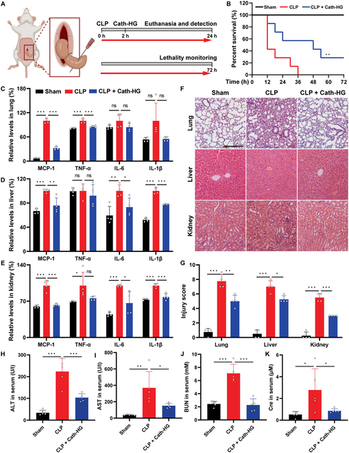
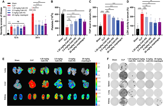
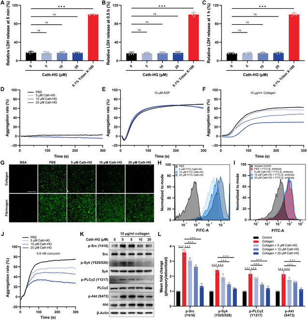
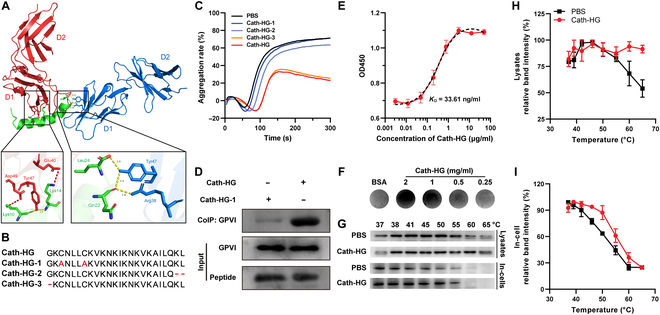
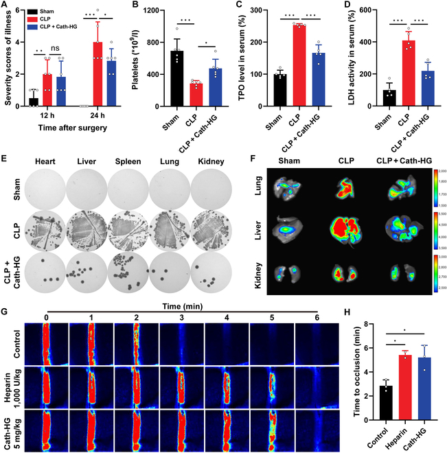
Platelet activation contributes to sepsis development, leading to microthrombosis and increased inflammation, which results in disseminated intravascular coagulation and multiple organ dysfunction. Although Cathelicidin can alleviate sepsis, its role in sepsis regulation remains largely unexplored. In this study, we identified Cath-HG, a novel Cathelicidin from Hylarana guentheri skin, and analyzed its structure using nuclear magnetic resonance spectroscopy. The modulatory effect of Cath-HG on the symptoms of mice with sepsis induced by cecal ligation and puncture was evaluated in vivo, and the platelet count, degree of organ damage, and microthrombosis were measured. The antiplatelet aggregation activity of Cath-HG was studied in vitro, and its target was verified. Finally, we further investigated whether Cath-HG could regulate thrombosis in vivo in a FeCl3 injury-induced carotid artery model. The results showed that Cath-HG exhibited an α-helical structure in sodium dodecyl sulfate solution and effectively reduced organ inflammation and damage, improving survival in septic mice. It alleviated sepsis-induced thrombocytopenia and microthrombosis. In vitro, Cath-HG specifically inhibited collagen-induced platelet aggregation and modulated glycoprotein VI (GPVI) signaling pathways. Dot blotting, enzyme-linked immunosorbent assay, and pull-down experiments confirmed GPVI as the target of Cath-HG. Molecular docking and amino acid residue truncations/mutations identified crucial sites of Cath-HG. These findings suggest that GPVI represents a promising therapeutic target for sepsis, and Cath-HG may serve as a potential treatment for sepsis-related thrombocytopenia and thrombotic events. Additionally, identifying Cath-HG as a GPVI inhibitor provides insights for developing novel antithrombotic therapies targeting platelet activation mediated by GPVI.


Conflict of interest statement

Competing interests: A patent was filed based on this work.

Figures

Fig. 1.
Fig. 1.
Identification and characterization of Cath-HG from H. guentheri. (A) The nucleotide sequence and predicted precursor sequence of Cath-HG. The presumed mature peptide is framed. The stop codon is represented by *. The signal peptide is gray shaded and is followed by a cathelin domain ending with KR residues (in red bold italics). The nucleotide and amino acid numbers are shown after the sequences. (B) Partial sequence similarity of Cath-HG precursor to homolog proteins from other amphibians (Ol, O. livida, AXR75914.1; Lc, Lithobates catesbeianus, QBC65431.1; Pn, Pelophylax nigromaculatus, QPL18198.1; Ny, Nanorana yunnanensis, AFX61592.1; Hr, Hoplobatrachus rugulosus, QQG31491.1; Rt, Rana temporaria, XP_040211152.1; Np, Nanorana parkeri, XP_018430778.1). Cathelin domain is indicated. The panel was generated with ESPript 3.0 (https://espript.ibcp.fr/ESPript/cgi-bin/ESPript.cgi). (C) Lowest–energy structure of Cath-HG with residue numbers and side chain. The secondary structure is represented in cartoon form, red indicates the α-helix, and yellow shows the Cys3–Cys7 disulfide bond that forms the N-terminal ring structure. The random coils are labeled with blue at the N- and the C-terminals. (D) Molecular surface of Cath-HG colored by electrostatic potential, with 2 colors describing the full range of positive (blue) and negative (red) charge across the surface.
Fig. 2.
Fig. 2.
Cath-HG protects mice from sepsis-induced multiple organ dysfunction. (A) Experimental design involving Cath-HG treatment (5 mg/kg) to CLP mice. (B) Survival rate of CLP-induced septic mice after Cath-HG treatment (n = 7). (C to E) The concentrations of proinflammatory cytokines in lungs, liver, and kidneys measured by ELISA (n = 4). (F) Representative images of histopathological analysis of lungs, liver, and kidneys stained with H&E (scale bar = 200 μm). (G) Injury scores of lungs, liver, and kidneys in each group (n = 4). (H to K) The levels of alanine aminotransferase (ALT), aspartate aminotransferase (AST), blood urea nitrogen (BUN), and creatinine (Cre) in the serum (n = 5). Data are expressed as mean ± SD. *P < 0.05; **P < 0.01; ***P < 0.001; ns, not significant.
Fig. 3.
Fig. 3.
Cath-HG attenuates excessive platelet aggregation in septic mice. (A) Severity scores of illness for each group at 12 and 24 h after CLP. Mice were intraperitoneally injected with Cath-HG (1.25, 2.5, and 5 mg/kg) or clopidogrel (20 mg/kg) 2 h after CLP (n = 4). (B to D) Platelet counts, TOP levels, and LDH activity in serum of mice in each group (n = 4). (E) Microvascular thrombosis in the lungs, liver, and kidneys detected by fluorescent microbeads through imaging. (F) Bacterial loads in the heart, liver, spleen, lung, and kidney. Data are expressed as mean ± SD. **P < 0.01; ***P < 0.001; ns, not significant.
Fig. 4.
Fig. 4.
Cath-HG inhibits collagen-induced platelet aggregation. (A to C) Release of LDH from washed platelets after Cath-HG (5, 10, and 20 μM) treatment for 5 min, 0.5 h, and 1 h (n = 5). (D) Platelet aggregation of PRP after Cath-HG (20 μM) treatment for 5 min. (E and F) Platelet aggregation of PRP induced by ADP (10 μM) and collagen (10 μg/ml) after Cath-HG (5, 10, and 20 μM) treatment for 5 min. (G) Spreading of platelets (green fluorescence) on collagen and fibrinogen (scale bar = 80 μm). (H) Flow cytometry analysis of Cath-HG (5, 10, and 20 μM) binding to platelets. (I) Flow cytometry analysis of the effect of Cath-HG (5, 10, and 20 μM) on the binding of platelet and β1 antibody. (J) Platelet aggregation of PRP induced by convulxin (0.6 nM) after Cath-HG (5, 10, and 20 μM) treatment for 5 min. (K) Immunoblot and (L) corresponding quantification analysis of phosphorylation of Src, Syk, PLCγ2, and Akt in washed platelets stimulated with collagen after 5-min treatment with or without Cath-HG (5, 10, and 20 μM). β-Actin was used as a loading control (n = 3). Data are expressed as mean ± SD. ***P < 0.001; ns, not significant.
Fig. 5.
Fig. 5.
Cath-HG directly targets to collagen receptor GPVI. (A) The 3D image of molecular docking showing Cath-HG (green) binding to the D1 domain of GPVI (red and blue). Lys10, Lys14, Leu24, and Gln22 of Cath-HG are predicted to interact with Arg38, Glu40, Try47, and Asp49 of GPVI. Red dotted lines indicate hydrophobic interactions, and yellow dotted lines indicate hydrogen bonds. (B) Sequence alignment of Cath-HG and its modified peptides. (C) Platelet aggregation of PRP induced by collagen (10 μg/ml) after Cath-HG or its modified peptides (20 μM) treatment for 5 min. (D) Pull-down analysis of Cath-HG selectively binding to GPVI. (E and F) Binding reaction between Cath-HG and GPVI. The platelet lysate was incubated with the indicated concentrations of Cath-HG immobilized in a 96-well plate or PVDF membranes before the contents of GPVI was detected by anti-GPVI primary antibody with corresponding ELISA (E) and dot blot (F) method, respectively (n = 3). (G to I) CETSA. The platelet or platelet lysate were incubated with Cath-HG (50 μM) at room temperature for 30 min and then the mixtures were heated for 3 min at indicated temperatures (37 to 65 °C) before the contents of GPVI were detected by Western blot (n = 3). Data are mean ± SD.
Fig. 6.
Fig. 6.
Cath-HG inhibits thrombus formation in vivo. Cath-HG (5 mg/kg) was intravenously injected into mice after 2 h of CLP, and the severity scores of illness (A), platelet count (B), TPO level (C), and LDH level (D) in the serum were determined (n = 5 to 6). (E) Lysogeny broth agar plate culture detecting bacterial load in the heart, liver, spleen, lungs, and kidneys. (F) Fluorescence imaging detecting microvascular thrombosis in the lungs, liver, and kidneys of CLP mice after intravenous injection of Cath-HG (5 mg/kg). (G) Representative images of carotid artery blood flow in FeCl3-treated mice. The mice received intravenous injections of 0.9% saline (w/v), heparin (1,000 U/kg), or Cath-HG (5 mg/kg) 30 min prior to injury and were subsequently monitored using a laser speckle flow imaging system. Red indicates blood flow, while blue-black areas indicate background. (H) Complete occlusion time of carotid arteries in FeCl3-injured mice after intravenous injection of samples (n = 3). Data are expressed as mean ± SD. *P < 0.05; **P < 0.01; ***P < 0.001; ns, not significant.

Similar articles

References

    1. Poll T, Shankar-Hari M, Wiersinga WJ. The immunology of sepsis. Immunity. 2021;54(11):2450–2464. - PubMed
    1. Du X, Zhang M, Zhou H, Wang W, Zhang C, Zhang L, Qu Y, Li W, Liu X, Zhao M, et al. . Decoy nanozymes enable multitarget blockade of proinflammatory cascades for the treatment of multi-drug-resistant bacterial sepsis. Research. 2022;2022:9767643. - PMC - PubMed
    1. Bao W, Xing H, Cao S, Long X, Liu H, Ma J, Guo F, Deng Z, Liu X. Neutrophils restrain sepsis associated coagulopathy via extracellular vesicles carrying superoxide dismutase 2 in a murine model of lipopolysaccharide induced sepsis. Nat Commun. 2022;13(1):4583. - PMC - PubMed
    1. Iba T, Levy JH, Warkentin TE, Thachil J, Poll T, Levi M, Scientific and Standardization Committee on DIC, and the Scientific and Standardization Committee on Perioperative and Critical Care of the International Society on Thrombosis and Haemostasis . Diagnosis and management of sepsis-induced coagulopathy and disseminated intravascular coagulation. J Thromb Haemost. 2019;17(11):1989–1994. - PubMed
    1. Iba T, Watanabe E, Umemura Y, Wada T, Hayashida K, Kushimoto S, Japanese Surviving Sepsis Campaign Guideline Working Group for disseminated intravascular coagulation, Wada H. Sepsis-associated disseminated intravascular coagulation and its differential diagnoses. J Intensive Care. 2019;7(1):32. - PMC - PubMed

LinkOut - more resources