

Impact of fucosyltransferase 1-mediated epidermal blood group antigen H on anti-inflammatory response in atopic dermatitis
- PMID: 38840912
- PMCID: PMC11151169
- DOI: 10.3389/fimmu.2024.1365430
Impact of fucosyltransferase 1-mediated epidermal blood group antigen H on anti-inflammatory response in atopic dermatitis
Abstract
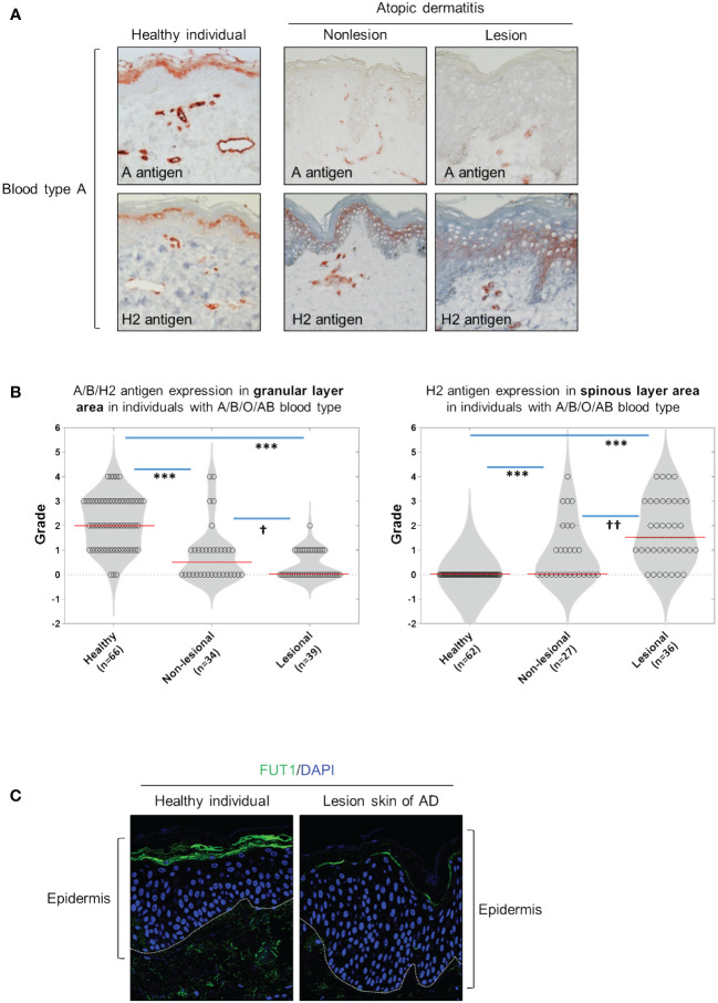
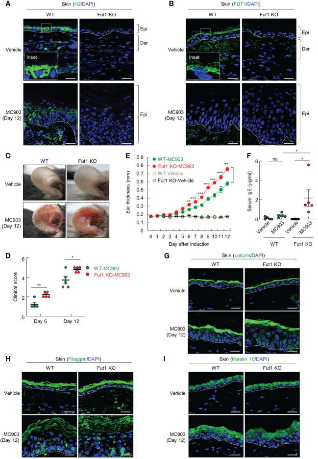
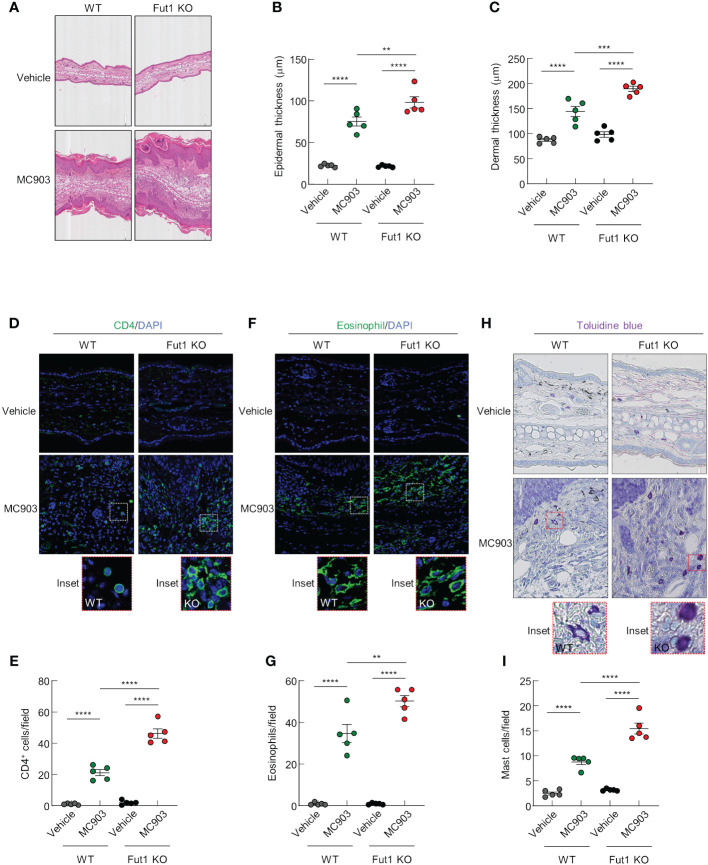
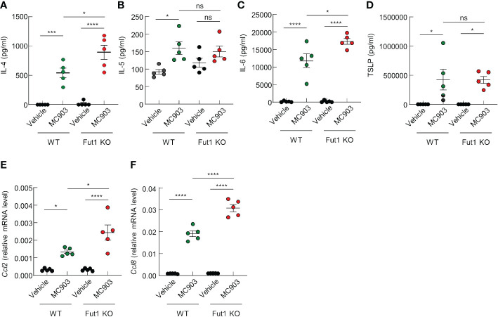
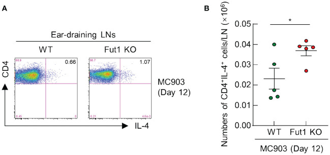
The presence of the blood group H2 antigen on the membrane of red blood cells determines blood type O in individuals and this H2 antigen serves as a precursor to the A and B antigens expressed in blood types A and B, respectively. However, the specific involvement of ABH antigens in skin diseases is unknown. Therefore, we aim to investigate the expression of ABH antigens in skin tissue of patients with atopic dermatitis (AD) and MC903-induced AD-like mice. We demonstrated that the expression of ABH antigen is primarily located in the granular and horny layers of the skin in healthy control individuals. However, in patients with AD, the expression of the ABH antigen was absent or diminished in these layers, while the H2 antigen expression increased in the spinous layers of the affected skin lesions. Then, we investigated the biological function of blood group H antigen mediated by fucosyltransferase 1 (Fut1) in the skin, utilizing an AD mouse model induced by MC903 in wild-type (WT) and Fut1-knockout mice. After the application of MC903, Fut1-deficient mice, with no H2 antigen expression on their skin, exhibited more severe clinical signs, increased ear swelling, and elevated serum IgE levels compared with those of WT mice. Additionally, the MC903-induced thickening of both the epidermis and dermis was more pronounced in Fut1-deficient mice than that in WT mice. Furthermore, Fut1-deficient mice showed a significantly higher production of interleukin-4 (IL-4) and IL-6 in skin lesions compared with that of their WT counterparts. The expression of chemokines, particularly Ccl2 and Ccl8, was notably higher in Fut1-deficient mice compared with those of WT mice. The infiltration of CD4+ T cells, eosinophils, and mast cells into the lesional skin was significantly elevated in Fut1-deficient mice compared with that in WT mice. These findings demonstrate the protective role of H2 antigen expression against AD-like inflammation and highlight its potential therapeutic impact on AD through the regulation of blood group antigens.
Keywords: ABH antigens; atopic dermatitis; chemokines; cytokines; fucosyltransferase 1.
Copyright © 2024 Li, Oh, Suh, Jin, Lee, Lee and Chung.
Conflict of interest statement
The authors declare that the research was conducted in the absence of any commercial or financial relationships that could be construed as a potential conflict of interest.
Figures
Similar articles
-
H Antigen expression modulates epidermal Keratinocyte Integrity and differentiation.Biol Res. 2024 Oct 18;57(1):72. doi: 10.1186/s40659-024-00541-x. Biol Res. 2024. PMID: 39420441 Free PMC article.
-
Fucosylation deficiency enhances imiquimod-induced psoriasis-like skin inflammation by promoting CXCL1 expression.Biochim Biophys Acta Mol Basis Dis. 2024 Feb;1870(2):166988. doi: 10.1016/j.bbadis.2023.166988. Epub 2023 Dec 7. Biochim Biophys Acta Mol Basis Dis. 2024. PMID: 38070583
-
Thymic stromal lymphopoietin-induced interleukin-17A is involved in the development of IgE-mediated atopic dermatitis-like skin lesions in mice.Immunology. 2015 Dec;146(4):568-81. doi: 10.1111/imm.12528. Epub 2015 Sep 24. Immunology. 2015. PMID: 26310839 Free PMC article.
-
The involvement of eosinophils in the patch test reaction to aeroallergens in atopic dermatitis: its relevance for the pathogenesis of atopic dermatitis.Clin Exp Allergy. 1993 Feb;23(2):97-109. doi: 10.1111/j.1365-2222.1993.tb00304.x. Clin Exp Allergy. 1993. PMID: 8448687 Review.
-
The Involvement of IL-34 in Inflammatory and Malignant Skin Diseases.J Invest Dermatol. 2025 Jun 16:S0022-202X(25)00490-7. doi: 10.1016/j.jid.2025.05.003. Online ahead of print. J Invest Dermatol. 2025. PMID: 40522246 Review.
Cited by
-
H Antigen expression modulates epidermal Keratinocyte Integrity and differentiation.Biol Res. 2024 Oct 18;57(1):72. doi: 10.1186/s40659-024-00541-x. Biol Res. 2024. PMID: 39420441 Free PMC article.
References
MeSH terms
Substances
LinkOut - more resources
Full Text Sources
Molecular Biology Databases
Research Materials