

Differences in IDO1+ dendritic cells and soluble CTLA-4 are associated with differential clinical responses to methotrexate treatment in rheumatoid arthritis
- PMID: 38840915
- PMCID: PMC11150726
- DOI: 10.3389/fimmu.2024.1352251
Differences in IDO1+ dendritic cells and soluble CTLA-4 are associated with differential clinical responses to methotrexate treatment in rheumatoid arthritis
Abstract
Objective: Antigen-presenting dendritic cells (DCs) and monocytes play an essential role in rheumatoid arthritis (RA) pathogenesis, however, their tolerogenic potential remains unclear. Herein, the tolerogenic profiles of DCs are characterized in treatment-naïve RA patients to determine their role to inflammatory arthritis management.
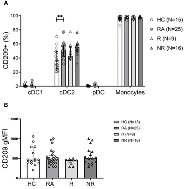
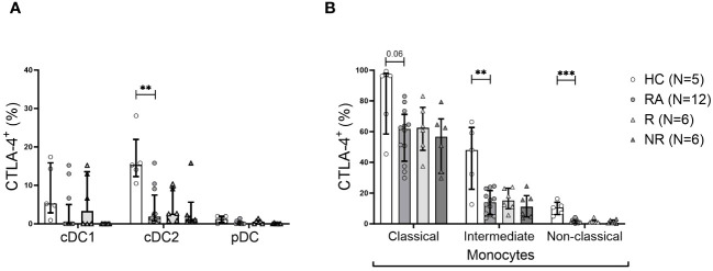
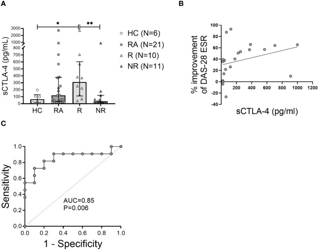
Methods: Thirty-six treatment-naïve RA patients were enrolled, of which 62% were non-responders to methotrexate (MTX) monotherapy based on disease activity score (DAS) after 6-months of therapy. DC and monocyte subset frequencies, activation (CD40, CD86, CD209 expression), and tolerogenic profile (intracellular indoleamine-2,3-dioxygenase [IDO1] and cytotoxic T lymphocyte antigen 4 [CTLA-4] expression) were examined in the baseline peripheral blood by multicolor flow-cytometry. Soluble CTLA-4 (sCTLA-4) levels in plasma were measured.
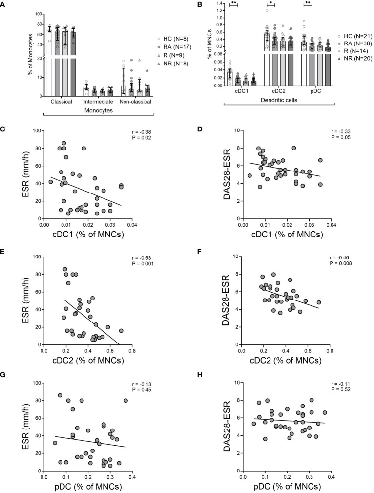
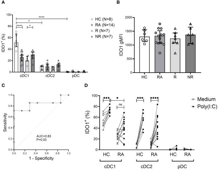
Results: DC subsets were decreased in RA compared to healthy controls (HC), and the frequency of conventional DCs (cDC) inversely correlated with inflammatory markers and improvement in disease activity. CD141+ cDC1s were the major IDO1-expressing cells. IDO1+cDC1s were reduced in RA patients compared to HC. The baseline frequency of IDO1+cDC1s inversely correlated with improvement in disease activity. CTLA-4 expression in CD1c+ cDC2s and monocytes was lower in RA patients compared to HC. Moreover, MTX-responders had a significantly lower frequency of IDO1+cDC1 cells and higher level of sCTLA-4 in the plasma compared to MTX non-responders. There was a strong predictive association of low IDO1+cDC1 cells, low sCTLA-4 and non-response to MTX.
Conclusions: Our findings reveal altered DC and monocytes immunophenotypes that are associated with RA pathology and treatment response. The frequencies of tolerogenic IDO1+cDC1s and the low level of sCTLA-4 are strongly associated with MTX non-responsiveness and therapeutic outcome. These results suggest that investigation of the association IDO1+cDC1 and sCTLA-4 with response to treatment may be more generalizable to other autoimmune diseases.
Keywords: CTLA-4; IDO1; dendritic cells; methotrexate; rheumatoid arthritis; therapy response; tolerogenic.
Copyright © 2024 Malik, Slauenwhite, McAlpine, Hanly, Marshall and Issekutz.
Conflict of interest statement
The authors declare that the research was conducted in the absence of any commercial or financial relationships that could be construed as a potential conflict of interest.
Figures
Similar articles
-
Phenotypic and Transcriptomic Analysis of Peripheral Blood Plasmacytoid and Conventional Dendritic Cells in Early Drug Naïve Rheumatoid Arthritis.Front Immunol. 2018 May 9;9:755. doi: 10.3389/fimmu.2018.00755. eCollection 2018. Front Immunol. 2018. PMID: 29867920 Free PMC article.
-
Association of a Type 2-Polarized T Cell Phenotype With Methotrexate Nonresponse in Patients With Rheumatoid Arthritis.Arthritis Rheumatol. 2020 Jul;72(7):1091-1102. doi: 10.1002/art.41223. Epub 2020 May 29. Arthritis Rheumatol. 2020. PMID: 32039570
-
Novel role of plasmacytoid dendritic cells in humans: induction of interleukin-10-producing Treg cells by plasmacytoid dendritic cells in patients with rheumatoid arthritis responding to therapy.Arthritis Rheum. 2010 Jan;62(1):53-63. doi: 10.1002/art.25037. Arthritis Rheum. 2010. PMID: 20039433
-
Pharmacomicrobiology of Methotrexate in Rheumatoid Arthritis: Gut Microbiome as Predictor of Therapeutic Response.Front Immunol. 2021 Dec 16;12:789334. doi: 10.3389/fimmu.2021.789334. eCollection 2021. Front Immunol. 2021. PMID: 34975886 Free PMC article. Review.
-
Dendritic cells as targets for therapy in rheumatoid arthritis.Nat Rev Rheumatol. 2009 Oct;5(10):566-71. doi: 10.1038/nrrheum.2009.185. Nat Rev Rheumatol. 2009. PMID: 19798032 Free PMC article. Review.
References
-
- Duong SQ, Crowson CS, Athreya A, Atkinson EJ, Davis JM, 3rd, Warrington KJ, et al. . Clinical predictors of response to methotrexate in patients with rheumatoid arthritis: a machine learning approach using clinical trial data. Arthritis Res Ther. (2022) 24:162. doi: 10.1186/s13075-022-02851-5 - DOI - PMC - PubMed
-
- Smolen JS, Landewe RBM, Bergstra SA, Kerschbaumer A, Sepriano A, Aletaha D, et al. . EULAR recommendations for the management of rheumatoid arthritis with synthetic and biological disease-modifying antirheumatic drugs: 2022 update. Ann Rheum Dis. (2023) 82:3–18. doi: 10.1136/ard-2022-223356corr1 - DOI - PubMed
MeSH terms
Substances
LinkOut - more resources
Full Text Sources
Medical
Research Materials