. 2024 May 22:15:1391968.
doi: 10.3389/fmicb.2024.1391968. eCollection 2024.

Carbonate precipitation and phosphate trapping by microbialite isolates from an alkaline insular lake (Bagno dell'Acqua, Pantelleria Island, Italy)

Affiliations

Carbonate precipitation and phosphate trapping by microbialite isolates from an alkaline insular lake (Bagno dell'Acqua, Pantelleria Island, Italy)

Cristina Mazzoni et al. Front Microbiol. .

Abstract

The Bagno dell'Acqua lake is characterized by CO2 emissions, alkaline waters (pH = 9) and Eh values which indicate strongly oxidizing conditions. A typical feature of the lake is the presence of actively growing microbialites rich in calcium carbonates and silica precipitates. Mineralogy, petrography and morphology analyses of the microbialites were coupled with the analysis of the microbial community, combining molecular and cultivation approaches. The DNA sequencing revealed distinct patterns of microbial diversity, showing pronounced differences between emerged and submerged microbialite, with the upper layer of emerged samples exhibiting the most distinctive composition, both in terms of prokaryotes and eukaryotes. In particular, the most representative phyla in the microbial community were Proteobacteria, Actinobacteriota, and Bacteroidota, while Cyanobacteria were present only with an average of 5%, with the highest concentration in the submerged intermediate layer (12%). The role of microorganisms in carbonate mineral formation was clearly demonstrated as most of the isolates were able to precipitate calcium carbonate and five of them were characterized at molecular level. Interestingly, when microbial isolates were cultivated only in filtered water, the precipitation of hazenite was observed (up to 85%), opening new prospective in P (phosphate) recovery from P depleted environments.

Keywords: alkaline lake; biomineralization; hazenite; microbialites; pantelleria; phosphate trapping.


Conflict of interest statement

The authors declare that the research was conducted in the absence of any commercial or financial relationships that could be construed as a potential conflict of interest.

Figures

Figure 1
Figure 1
(A) Map of Italy with focus on the island of Pantelleria; (B) Satellite image reporting sampling locations labeled as 1 (emerged microbialite) and 2 (submerged microbialite); (C) emerged microbialite sample with the three analyzed layers (1-W, 1-G, 1-B); (D) submerged microbialite sample with the three analyzed layers (2-W, 2-G, 2-B).
Figure 2
Figure 2
SEM images representative of the three differently colored layers. (A) In the W layer a dense EPS network with abundant diatom frustules and aggregates of silicate (Mg-smectite) and carbonate minerals is observable. (A) detrital feldspar occurs in the lower left. (B) In the G layer the EPS matrix in association with smectite aggregates is diffuse. Diatom frustules, many of them partially corroded are scarcely represented. (C) In the B layer aggregates of carbonate minerals along with smectite coat detrital minerals. Note in the red circle the presence of rosette-like clusters of calcium-carbonate minerals (possibly aragonite). Kfs, K-feldspar; EPS, Extracellular Polymeric Substances.
Figure 3
Figure 3
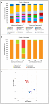
Relative abundance (%) of the most prevalent phyla of (A) Bacteria and (B) Archaea. The graph shows only phyla which contributed more than 1.5% to the total bacterial community in at least one sample. The abundance of the remaining phyla was summed and labeled as “other”. (C) Principal Component Analysis (PCA) performed on the phyla of Bacteria and Archaea. Identification of samples with similar microbial communities using PCA. Each point represents the microbial community in each specific sample. The distance between points on the plot corresponds to similarity; closer points refer to samples with highly similar microbial communities (source: Rstudio, version 4.3.1).
Figure 4
Figure 4
Crystal formation kinetics. Cells from saturated culture of Stappia sp. 3bis2 strain were spotted onto B4CaCl2 pH8, B4CaLact pH8 and B4CaLact pH6 plates, incubated at 28°C and observed by optical microscopy after 5, 7 and 11 days. Scale bar: 100 μm in B4CaLact pH6, 5 Days, 200 μm in all the other panels.
Figure 5
Figure 5
SEM images representative of carbonate-crystals from strains (A) Stappia sp. 3bis2; (B) Bacillus sp. 3bis3; (C) Sutcliffiella sp. 3bis5; (D) Metabacillus sp. 3bis7; (E) Bacillus sp. 3bis8 grown in B4CaLactate medium for 16 (3bis2) and 24 days (3bis3, 5, 7, and 8). (F) is an enlargement of (C) showing bacterial cells calcification.
Figure 6
Figure 6
Stappia sp. 3bis2 cells formed hazenite when deposited onto B4LW and incubate for 35 days at 28°C. (A) Stereomicroscope image; scale bar: 1 mm; (B) Optical microscopy image showed crystals in the bar or aciculate forms; scale bar 200 μm. (C) Field-emission scanning electron microscopy (FE-SEM) of crystals, red circle represents the point where EDS was applied; (D) EDS.
Figure 7
Figure 7
All the isolates can precipitate hazenite after 35 days of incubation. (A) Stappia sp. 3bis2; (B) Bacillus sp. 3bis3; (C) Sutcliffiella sp. 3bis5; (D) Metabacillus sp. 3bis7; grown in B4LW for 35 days (B, C, D); (E) Bacillus sp. 3bis8 grown in B4LW for 100 days; (F, G) represent EDS analysis of regions indicated by the red squares in (C, D), respectively.
Figure 8
Figure 8
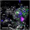
CLSM combined images showing the spatial distribution of Bacteria belonging to the class Bacilli (phylum Firmicutes (green) and other DAPI stained cells (blue) identified by CARD-FISH in the deep layers of the natural microbialite (1-B). Autofluorescent cells appear in red. The hybridized bacterial cells were excited with the 488 nm line of an Ar laser (excitation) and observed in the green channel from 500 to 530 nm (emission). Mineral crystals were visualized by their reflection signal (405 nm line of a diode laser) and appear of gray color. Bar = 20 μm.

References

    1. Abdel-Azeem A. M., Salem F. M., Abdel-Azeem M. A., Nafady N. A., Mohesien M. T., Soliman E. A. (2016). “Biodiversity of the genus Aspergillus in different habitats,” in New and Future Developments in Microbial Biotechnology and Bioengineering (Amsterdam: Elsevier; ), 3–28.
    1. Albertsen M., Karst S. M., Ziegler A. S., Kirkegaard R. H., Nielsen P. H. (2015). Back to basics–the influence of DNA extraction and primer choice on phylogenetic analysis of activated sludge communities. PLoS ONE 10:e0132783. 10.1371/journal.pone.0132783 - DOI - PMC - PubMed
    1. Amalfitano S., Fazi S. (2008). Recovery and quantification of bacterial cells associated with streambed sediments. J. Microbiol. Methods 75, 237–243. 10.1016/j.mimet.2008.06.004 - DOI - PubMed
    1. Apprill A., McNally S., Parsons R., Weber L. (2015). Minor revision to V4 region SSU rRNA 806R gene primer greatly increases detection of SAR11 bacterioplankton. Aquatic Microb. Ecol. 75, 129–137. 10.3354/ame01753 - DOI
    1. Azua-Bustos A., Fairén A. G., González-Silva C., Prieto-Ballesteros O., Carrizo D., Sánchez-García L., Rampe E. (2023). Dark microbiome and extremely low organics in Atacama fossil delta unveil Mars life detection limits. Nat. Commun. 14:808. 10.1038/s41467-023-36172-1 - DOI - PMC - PubMed

LinkOut - more resources