

Immune-related gene methylation prognostic instrument for stratification and targeted treatment of ovarian cancer patients toward advanced 3PM approach
- PMID: 38841623
- PMCID: PMC11148001
- DOI: 10.1007/s13167-024-00359-3
Immune-related gene methylation prognostic instrument for stratification and targeted treatment of ovarian cancer patients toward advanced 3PM approach
Abstract
Background: DNA methylation is an important mechanism in epigenetics, which can change the transcription ability of genes and is closely related to the pathogenesis of ovarian cancer (OC). We hypothesize that DNA methylation is significantly different in OCs compared to controls. Specific DNA methylation status can be used as a biomarker of OC, and targeted drugs targeting these methylation patterns and DNA methyltransferase may have better therapeutic effects. Studying the key DNA methylation sites of immune-related genes (IRGs) in OC patients and studying the effects of these methylation sites on the immune microenvironment may provide a new method for further exploring the pathogenesis of OC, realizing early detection and effective monitoring of OC, identifying effective biomarkers of DNA methylation subtypes and drug targets, improving the efficacy of targeted drugs or overcoming drug resistance, and better applying it to predictive diagnosis, prevention, and personalized medicine (PPPM; 3PM) of OC.
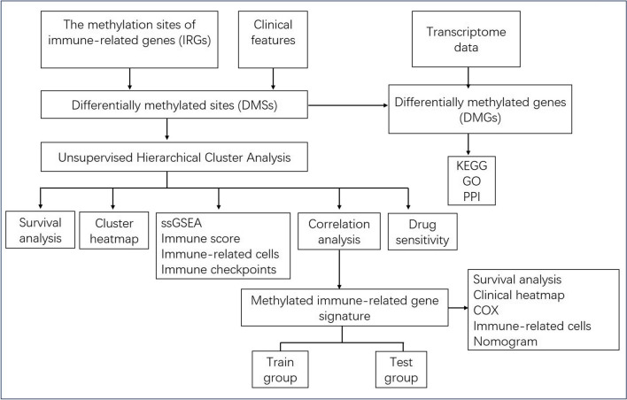
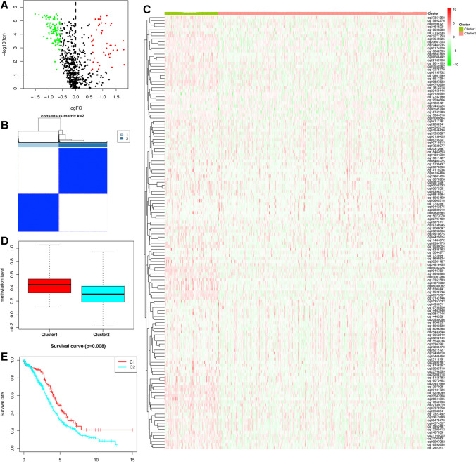
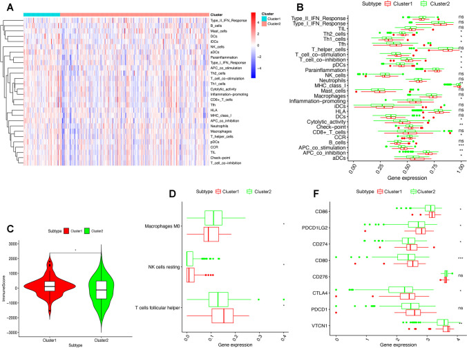
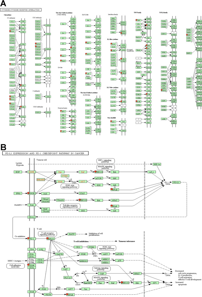
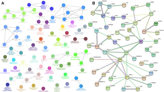
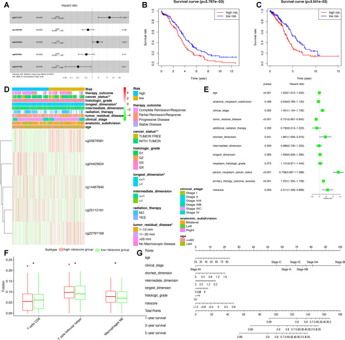
Method: Hypermethylated subtypes (cluster 1) and hypomethylated subtypes (cluster 2) were established in OCs based on the abundance of different methylation sites in IRGs. The differences in immune score, immune checkpoints, immune cells, and overall survival were analyzed between different methylation subtypes in OC samples. The significant pathways, gene ontology (GO), and protein-protein interaction (PPI) network of the identified methylation sites in IRGs were enriched. In addition, the immune-related methylation signature was constructed with multiple regression analysis. A methylation site model based on IRGs was constructed and verified.
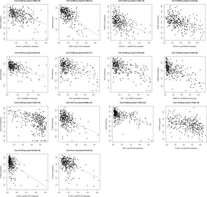
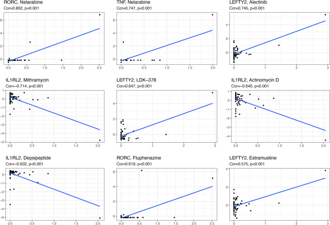
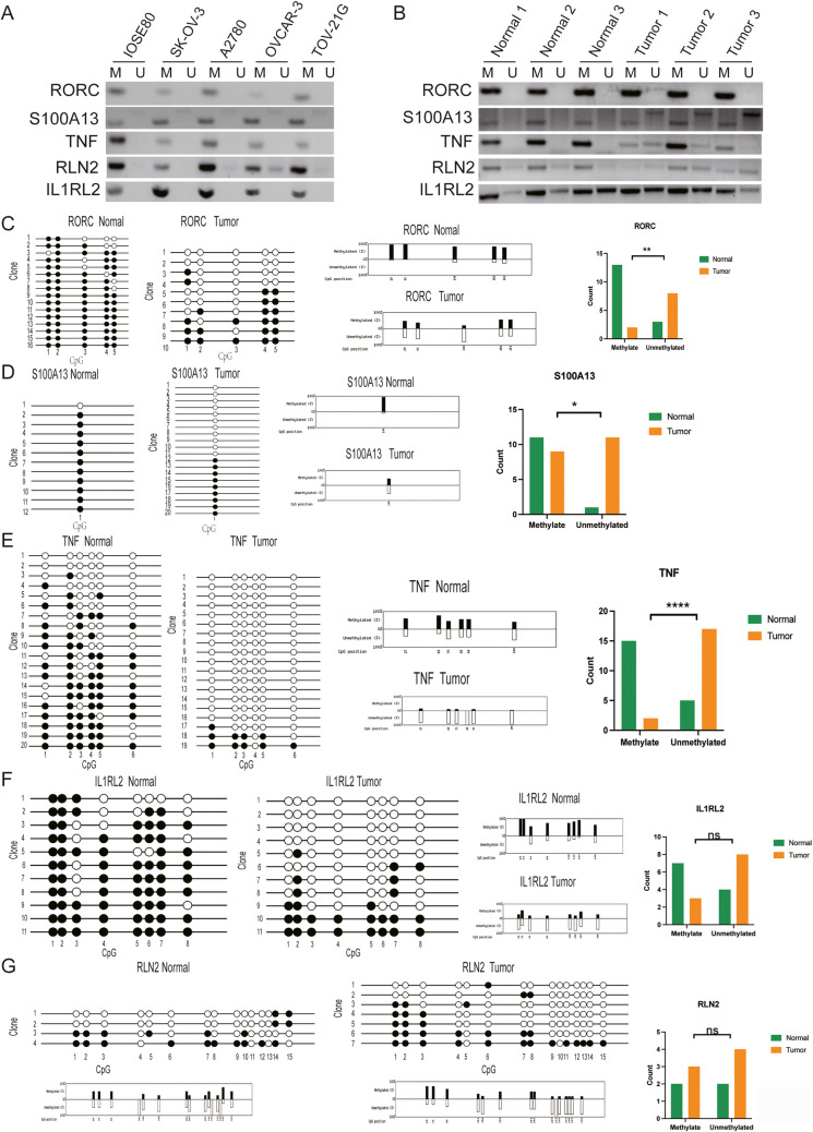
Results: A total of 120 IRGs with 142 differentially methylated sites (DMSs) were identified. The DMSs were clustered into a high-level methylation group (cluster 1) and a low-level methylation group (cluster 2). The significant pathways and GO analysis showed many immune-related and cancer-associated enrichments. A methylation site signature based on IRGs was constructed, including RORC|cg25112191, S100A13|cg14467840, TNF|cg04425624, RLN2|cg03679581, and IL1RL2|cg22797169. The methylation sites of all five genes showed hypomethylation in OC, and there were statistically significant differences among RORC|cg25112191, S100A13|cg14467840, and TNF|cg04425624 (p < 0.05). This prognostic model based on low-level methylation and high-level methylation groups was significantly linked to the immune microenvironment as well as overall survival in OC.
Conclusions: This study provided different methylation subtypes for OC patients according to the methylation sites of IRGs. In addition, it helps establish a relationship between methylation and the immune microenvironment, which showed specific differences in biological signaling pathways, genomic changes, and immune mechanisms within the two subgroups. These data provide ones to deeply understand the mechanism of immune-related methylation genes on the occurrence and development of OC. The methylation-site signature is also to establish new possibilities for OC therapy. These data are a precious resource for stratification and targeted treatment of OC patients toward an advanced 3PM approach.
Supplementary information: The online version contains supplementary material available at 10.1007/s13167-024-00359-3.
Keywords: Biomarker; DNA methylation; Differentially methylated sites; Immune-related genes; Methylation subtypes; Methylation-site signature; Ovarian cancer; Overall survival; Patient stratification; Risk score; Targeted treatment; Tumor immune microenvironment; Predictive preventive personalized medicine (PPPM; 3PM).
© The Author(s), under exclusive licence to European Association for Predictive, Preventive and Personalised Medicine (EPMA) 2024. Springer Nature or its licensor (e.g. a society or other partner) holds exclusive rights to this article under a publishing agreement with the author(s) or other rightsholder(s); author self-archiving of the accepted manuscript version of this article is solely governed by the terms of such publishing agreement and applicable law.
Conflict of interest statement
Competing interestsThe authors declare no competing interests.
Figures
Similar articles
-
Clinically relevant immune subtypes based on alternative splicing landscape of immune-related genes for lung cancer advanced PPPM approach.EPMA J. 2024 May 24;15(2):345-373. doi: 10.1007/s13167-024-00366-4. eCollection 2024 Jun. EPMA J. 2024. PMID: 38841624 Free PMC article.
-
Clinically relevant stratification of lung squamous carcinoma patients based on ubiquitinated proteasome genes for 3P medical approach.EPMA J. 2024 Mar 4;15(1):67-97. doi: 10.1007/s13167-024-00352-w. eCollection 2024 Mar. EPMA J. 2024. PMID: 38463626 Free PMC article.
-
Quantitative phosphoproteomics reveals molecular pathway network alterations in human early-stage primary hepatic carcinomas: potential for 3P medical approach.EPMA J. 2023 Aug 10;14(3):477-502. doi: 10.1007/s13167-023-00335-3. eCollection 2023 Sep. EPMA J. 2023. PMID: 37605650 Free PMC article.
-
Nitroproteomics is instrumental for stratification and targeted treatments of astrocytoma patients: expert recommendations for advanced 3PM approach with improved individual outcomes.EPMA J. 2023 Dec 6;14(4):673-696. doi: 10.1007/s13167-023-00348-y. eCollection 2023 Dec. EPMA J. 2023. PMID: 38094577 Free PMC article. Review.
-
Energy metabolism as the hub of advanced non-small cell lung cancer management: a comprehensive view in the framework of predictive, preventive, and personalized medicine.EPMA J. 2024 Apr 8;15(2):289-319. doi: 10.1007/s13167-024-00357-5. eCollection 2024 Jun. EPMA J. 2024. PMID: 38841622 Free PMC article. Review.
Cited by
-
Multi-omics decipher the immune microenvironment and unveil therapeutic strategies for postoperative ovarian cancer patients.Transl Cancer Res. 2024 Nov 30;13(11):6028-6044. doi: 10.21037/tcr-24-656. Epub 2024 Nov 21. Transl Cancer Res. 2024. PMID: 39697734 Free PMC article.
-
Artificial intelligence in ovarian cancer drug resistance advanced 3PM approach: subtype classification and prognostic modeling.EPMA J. 2024 Jul 13;15(3):525-544. doi: 10.1007/s13167-024-00374-4. eCollection 2024 Sep. EPMA J. 2024. PMID: 39239109
-
Pharmacoproteomics reveals energy metabolism pathways as therapeutic targets of ivermectin in ovarian cancer toward 3P medical approaches.EPMA J. 2024 Nov 25;15(4):711-737. doi: 10.1007/s13167-024-00385-1. eCollection 2024 Dec. EPMA J. 2024. PMID: 39635022
References
-
- Roett MA, Evans P. Ovarian cancer: an overview. Am Fam Physician. 2009;80(6):609–616. - PubMed
LinkOut - more resources
Full Text Sources
