. 2024 Jul;38(7):506-517.
doi: 10.1177/15459683241257523. Epub 2024 Jun 6.

Cross-Frequency Coupling as a Biomarker for Early Stroke Recovery

Affiliations

Cross-Frequency Coupling as a Biomarker for Early Stroke Recovery

Jasper I Mark et al. Neurorehabil Neural Repair. 2024 Jul.

Abstract

Background: The application of neuroimaging-based biomarkers in stroke has enriched our understanding of post-stroke recovery mechanisms, including alterations in functional connectivity based on synchronous oscillatory activity across various cortical regions. Phase-amplitude coupling, a type of cross-frequency coupling, may provide additional mechanistic insight.

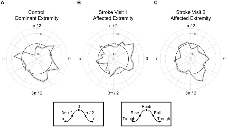
Objective: To determine how the phase of prefrontal cortex delta (1-3 Hz) oscillatory activity mediates the amplitude of motor cortex beta (13-20 Hz) oscillations in individual's early post-stroke.

Methods: Participants admitted to an inpatient rehabilitation facility completed resting and task-based EEG recordings and motor assessments around the time of admission and discharge along with structural neuroimaging. Unimpaired controls completed EEG procedures during a single visit. Mixed-effects linear models were performed to assess within- and between-group differences in delta-beta prefrontomotor coupling. Associations between coupling and motor status and injury were also determined.

Results: Thirty individuals with stroke and 17 unimpaired controls participated. Coupling was greater during task versus rest conditions for all participants. Though coupling during affected extremity task performance decreased during hospitalization, coupling remained elevated at discharge compared to controls. Greater baseline coupling was associated with better motor status at admission and discharge and positively related to motor recovery. Coupling demonstrated both positive and negative associations with injury involving measures of lesion volume and overlap injury to anterior thalamic radiation, respectively.

Conclusions: This work highlights the utility of prefrontomotor cross-frequency coupling as a potential motor status and recovery biomarker in stroke. The frequency- and region-specific neurocircuitry featured in this work may also facilitate novel treatment strategies in stroke.

Keywords: biomarker; connectivity; neuroimaging; rehabilitation; stroke.


Conflict of interest statement

Declaration of Conflicting InterestsThe author(s) declared the following potential conflicts of interest with respect to the research, authorship, and/or publication of this article: JM reports stock holdings in Impulse Wellness LLC. FF is the lead inventory of IP filed on the topics of non-invasive brain stimulation by UNC. FF has received honoraria from the following entities in the last 12 months: Electromedical Products International, Academic Press, and Insel Spital. All other authors have no competing interests to disclose.

Figures

Figure 1.
Figure 1.
Enhancement of PFC-M1 DB-PAC during task performance. Participants with stroke and controls demonstrated greater DB-PAC during the task (collapsed across extremity) versus rest condition. *P < .05. **P < .01. ***P < .001.
Figure 2.
Figure 2.
Enhanced ipsilesional PFC-M1 DB-PAC during task performance with the affected extremity. Individuals with stroke demonstrated significantly greater coupling during task performance with their affected versus less-affected extremity at visits 1 and 2 despite a significant decrease in coupling during affected extremity performance between visits. *P < .05. **P < .01. ***P < .001.
Figure 3.
Figure 3.
Stroke shifts M1 beta amplitude to the rise of PFC delta phase. Rose plots of PFC delta phase coupled to M1 beta amplitude (black line) in (A) control participants during task performance with the dominant extremity, (B) individuals with stroke during task performance with the affected extremity at visit 1 and visit 2 (C). A shift in phase-dependency from the peak of the delta PFC phase to the rise of the delta PFC phase occurred between visits 1 and 2. Grey shading around the black line indicates standard error. Peak and trough values in radians provide additional orientation.
Figure 4.
Figure 4.
Cluster analysis of prefrontal delta and scalp beta coupling. (A) Topographical plots demonstrating coupling between delta phase of electrodes overlying PFC (outlined box) and scalp beta amplitude. Electrodes showing a significant Group effect (Bonferroni-corrected P = .00026) are plotted for each visit. Darker red color reflects greater coupling. (B) Graphical presentation of the clustered F-statistic for the Group and Time interaction across electrodes triangulated to 10 to 20 neighbors presented. Larger electrodes reflect a greater interaction. Coupling is greatest in the ipsilesional hemisphere during affected extremity task performance (B, middle). During non-dominant and less-affected extremity performance, the interaction is greater in clustered electrodes overlying the contralesional hemisphere (B, bottom). L indicates the left or ipsilesional hemisphere, and R indicates the right or contralesional hemisphere.
Figure 5.
Figure 5.
Proposed conceptual model of a PFC-M1 DB-PAC network. Delta oscillatory activity from prefrontal cortex (PFC, purple) modulates beta oscillatory activity from nearby primary motor cortex (M1, blue) through (A) functional connections (dotted arrow) and (B) structural connections (solid arrow) via white matter fiber tracts such as the anterior thalamic radiation (ATR, green). (C) Smaller lesion volume and greater ATR overlap injury (red) are associated with less coupling. (D) Larger lesion volume and less ATR overlap injury are associated with greater coupling.

References

    1. Cramer SC. Repairing the human brain after stroke: I. Mechanisms of spontaneous recovery. Ann Neurol. 2008;63(3):272-287. - PubMed
    1. Cassidy JM, Tran G, Quinlan EB, Cramer SC. Neuroimaging identifies patients most likely to respond to a restorative stroke therapy. Stroke. 2018;49(2):433-438. - PMC - PubMed
    1. Tedesco Triccas L, Meyer S, Mantini D, et al. A systematic review investigating the relationship of electroencephalography and magnetoencephalography measurements with sensorimotor upper limb impairments after stroke. J Neurosci Methods. 2019;311:318-330. - PubMed
    1. Kawano T, Hattori N, Uno Y, et al. Association between aphasia severity and brain network alterations after stroke assessed using the electroencephalographic phase synchrony index. Sci Rep. 2021;11(1):12469. - PMC - PubMed
    1. Cassidy JM, Wodeyar A, Srinivasan R, Cramer SC. Coherent neural oscillations inform early stroke motor recovery. Hum Brain Mapp. 2021;42:5636-5647. - PMC - PubMed