

Effects of fixation and demineralization on histomorphology and DNA amplification of canine bone marrow
- PMID: 38842072
- PMCID: PMC11538782
- DOI: 10.1177/03009858241257920
Effects of fixation and demineralization on histomorphology and DNA amplification of canine bone marrow
Abstract
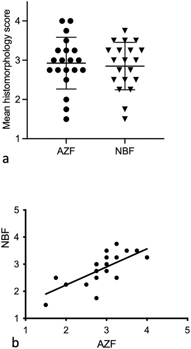
Fixation and demineralization protocols for bone marrow (BM) across diagnostic laboratories are not standardized. How different protocols affect histomorphology and DNA amplification is incompletely understood. In this study, 2 fixatives and 3 demineralization methods were tested on canine BM samples. Twenty replicate sternal samples obtained within 24 hours of death were fixed overnight in either acetic acid-zinc-formalin (AZF) or 10% neutral-buffered formalin (NBF) and demineralized with formic acid for 12 hours. Another 53 samples were fixed in AZF and demineralized with hydrochloric acid for 1-hour, formic acid for 12 hours, or ethylenediamine tetraacetic acid (EDTA) for 24 hours. Histologic sections were scored by 4 raters as of insufficient, marginal, good, or excellent quality. In addition, DNA samples extracted from sections treated with the different fixation and demineralization methods were amplified with 3 sets of primers to conserved regions of T cell receptor gamma and immunoglobulin heavy chain genes. Amplification efficiency was graded based on review of capillary electrophoretograms. There was no significant difference in the histomorphology scores of sections fixed in AZF or NBF. However, EDTA-based demineralization yielded higher histomorphology scores than demineralization with hydrochloric or formic acid, whereas formic acid resulted in higher scores than hydrochloric acid. Demineralization with EDTA yielded DNA amplification in 29 of 36 (81%) samples, whereas demineralization with either acid yielded amplification in only 2 of 72 (3%) samples. Although slightly more time-consuming and labor-intensive, tissue demineralization with EDTA results in superior morphology and is critical for polymerase chain reaction (PCR) amplification with the DNA extraction method described in this article.
Keywords: EDTA; PARR; bone; clonality; decalcification; dog; formalin; formic acid; hematopoietic tissue; hydrochloric acid; lymphocyte antigen receptor genes.
Conflict of interest statement
Declaration of Conflicting InterestsThe author(s) declared no potential conflicts of interest with respect to the research, authorship, and/or publication of this article.
Figures


References
-
- Bonds LA, Barnes P, Foucar K, et al. Acetic acid-zinc-formalin: a safe alternative to B-5 fixative. Am J Clin Pathol. 2005;124(2):205–211. - PubMed
-
- Brown RSD, Edwards J, Bartlett JW, et al. Routine acid decalcification of bone marrow samples can preserve DNA for FISH and CGH studies in metastatic prostate cancer. 2002;50(1):113–115. - PubMed
-
- Callis G, Sterchi D. Decalcification of bone: literature review and practical study of various decalcifying agents, methods, and their effects on bone histology. J Histotechnol. 1998;21(1):49–58.
-
- Castania VA, de Souza da Silveira JW, Issy AC, et al. Advantages of a combined method of decalcification compared to EDTA. Microsc Res Tech. 2015;78(2):111–118. - PubMed
