Amentoflavone reverses epithelial-mesenchymal transition in hepatocellular carcinoma cells by targeting p53 signalling pathway axis
- PMID: 38842135
- PMCID: PMC11154840
- DOI: 10.1111/jcmm.18442
Amentoflavone reverses epithelial-mesenchymal transition in hepatocellular carcinoma cells by targeting p53 signalling pathway axis
Abstract
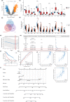
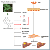
Epithelial-mesenchymal transition (EMT) and its reversal process are important potential mechanisms in the development of HCC. Selaginella doederleinii Hieron is widely used in Traditional Chinese Medicine for the treatment of various tumours and Amentoflavone is its main active ingredient. This study investigates the mechanism of action of Amentoflavone on EMT in hepatocellular carcinoma from the perspective of bioinformatics and network pharmacology. Bioinformatics was used to screen Amentoflavone-regulated EMT genes that are closely related to the prognosis of HCC, and a molecular prediction model was established to assess the prognosis of HCC. The network pharmacology was used to predict the pathway axis regulated by Amentoflavone. Molecular docking of Amentoflavone with corresponding targets was performed. Detection and evaluation of the effects of Amentoflavone on cell proliferation, migration, invasion and apoptosis by CCK-8 kit, wound healing assay, Transwell assay and annexin V-FITC/propidium iodide staining. Eventually three core genes were screened, inculding NR1I2, CDK1 and CHEK1. A total of 590 GO enrichment entries were obtained, and five enrichment results were obtained by KEGG pathway analysis. Genes were mainly enriched in the p53 signalling pathway. The outcomes derived from both the wound healing assay and Transwell assay demonstrated significant inhibition of migration and invasion in HCC cells upon exposure to different concentrations of Amentoflavone. The results of Annexin V-FITC/PI staining assay showed that different concentrations of Amentoflavone induces apoptosis in HCC cells. This study revealed that the mechanism of Amentoflavone reverses EMT in hepatocellular carcinoma, possibly by inhibiting the expression of core genes and blocking the p53 signalling pathway axis to inhibit the migration and invasion of HCC cells.
Keywords: Amentoflavone; bioinformatics; epithelial‐mesenchymal transition; hepatocellular carcinoma; network pharmacology; p53 signalling pathway axis.
© 2024 The Author(s). Journal of Cellular and Molecular Medicine published by Foundation for Cellular and Molecular Medicine and John Wiley & Sons Ltd.
Conflict of interest statement
The authors declare that the research was conducted in the absence of any commercial or financial relationships that could be construed as a potential conflict of interest.
Figures


Similar articles
-
Dendrobium nobile active ingredient Dendrobin A against hepatocellular carcinoma via inhibiting nuclear factor kappa-B signaling.Biomed Pharmacother. 2024 Aug;177:117013. doi: 10.1016/j.biopha.2024.117013. Epub 2024 Jun 19. Biomed Pharmacother. 2024. PMID: 38901205
-
Amentoflavone Inhibits ERK-modulated Tumor Progression in Hepatocellular Carcinoma In Vitro.In Vivo. 2018 May-Jun;32(3):549-554. doi: 10.21873/invivo.11274. In Vivo. 2018. PMID: 29695559 Free PMC article.
-
Mechanism of Dahuang-Dangshen drug pairs in the treatment of HCC based on network pharmacology, bioinformatics, molecular docking and experimental verification.Med Oncol. 2025 Apr 22;42(5):174. doi: 10.1007/s12032-025-02738-w. Med Oncol. 2025. PMID: 40261593
-
Wnt Signaling in Hepatocellular Carcinoma: Biological Mechanisms and Therapeutic Opportunities.Cells. 2024 Dec 2;13(23):1990. doi: 10.3390/cells13231990. Cells. 2024. PMID: 39682738 Free PMC article. Review.
-
Ribosomal proteins in hepatocellular carcinoma: mysterious but promising.Cell Biosci. 2024 Nov 1;14(1):133. doi: 10.1186/s13578-024-01316-3. Cell Biosci. 2024. PMID: 39487553 Free PMC article. Review.
Cited by
-
Amentoflavone protects against cisplatin-induced acute kidney injury by modulating Nrf2-mediated oxidative stress and ferroptosis and partially by activating Nrf2-dependent PANoptosis.Front Pharmacol. 2025 Mar 5;16:1508047. doi: 10.3389/fphar.2025.1508047. eCollection 2025. Front Pharmacol. 2025. PMID: 40110131 Free PMC article.
-
The multi-target mechanism of action of Selaginella doederleinii Hieron in the treatment of nasopharyngeal carcinoma: a network pharmacology and multi-omics analysis.Sci Rep. 2025 Jan 2;15(1):159. doi: 10.1038/s41598-024-83921-3. Sci Rep. 2025. PMID: 39747499 Free PMC article.
-
Potential mechanism of Camellia luteoflora against colon adenocarcinoma: An integration of network pharmacology and molecular docking.World J Gastrointest Oncol. 2025 Jun 15;17(6):105782. doi: 10.4251/wjgo.v17.i6.105782. World J Gastrointest Oncol. 2025. PMID: 40547172 Free PMC article.
References
-
- Sung H, Ferlay J, Siegel RL, et al. Global cancer statistics 2020: GLOBOCAN estimates of incidence and mortality worldwide for 36 cancers in 185 countries. CA Cancer J Clin. 2021;71(3):209‐249. - PubMed
-
- Yang R, Gao W, Wang Z, et al. Polyphyllin I induced ferroptosis to suppress the progression of hepatocellular carcinoma through activation of the mitochondrial dysfunction via Nrf2/HO‐1/GPX4 axis. Phytomedicine. 2024;122:155135. - PubMed
-
- Chen W, Zheng R, Zhang S, et al. Cancer incidence and mortality in China, 2013. Cancer Lett. 2017;401:63‐71. - PubMed
-
- Llovet JM, Castet F, Heikenwalder M, et al. Immunotherapies for hepatocellular carcinoma. Nat Rev Clin Oncol. 2022;19(3):151‐172. - PubMed
MeSH terms
Substances
Grants and funding
LinkOut - more resources
Full Text Sources
Medical
Research Materials
Miscellaneous

