Review
. 2024 Jul 10;124(13):8014-8129.
doi: 10.1021/acs.chemrev.3c00902. Epub 2024 Jun 6.

Condensed Matter Systems Exposed to Radiation: Multiscale Theory, Simulations, and Experiment

Affiliations
Review

Condensed Matter Systems Exposed to Radiation: Multiscale Theory, Simulations, and Experiment

Andrey V Solov'yov et al. Chem Rev. .

Abstract

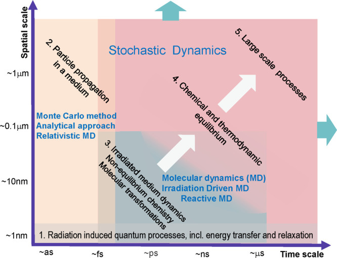
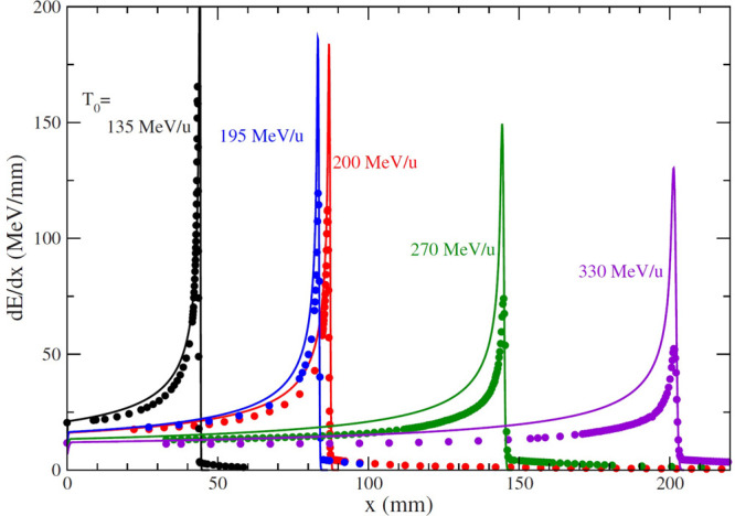
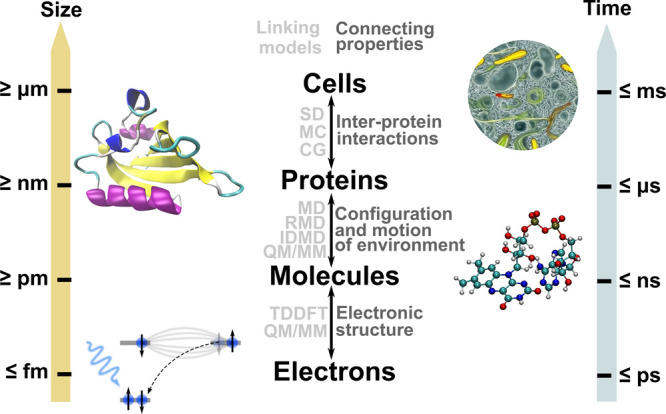
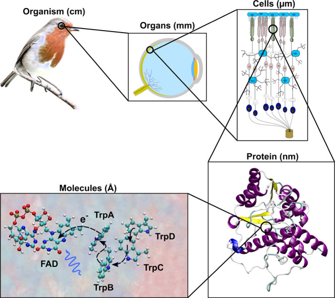
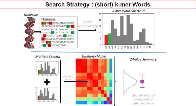
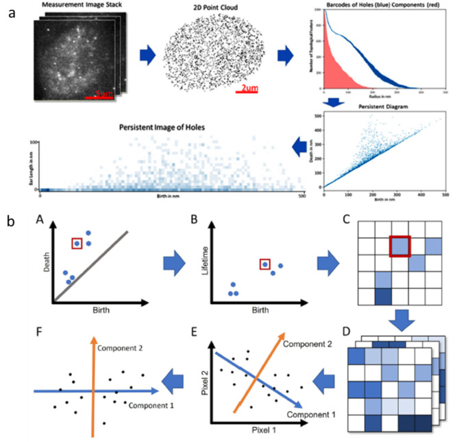
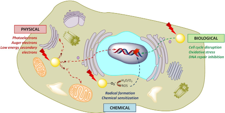
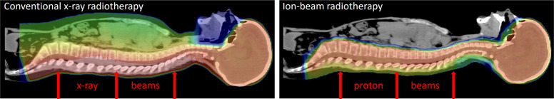
This roadmap reviews the new, highly interdisciplinary research field studying the behavior of condensed matter systems exposed to radiation. The Review highlights several recent advances in the field and provides a roadmap for the development of the field over the next decade. Condensed matter systems exposed to radiation can be inorganic, organic, or biological, finite or infinite, composed of different molecular species or materials, exist in different phases, and operate under different thermodynamic conditions. Many of the key phenomena related to the behavior of irradiated systems are very similar and can be understood based on the same fundamental theoretical principles and computational approaches. The multiscale nature of such phenomena requires the quantitative description of the radiation-induced effects occurring at different spatial and temporal scales, ranging from the atomic to the macroscopic, and the interlinks between such descriptions. The multiscale nature of the effects and the similarity of their manifestation in systems of different origins necessarily bring together different disciplines, such as physics, chemistry, biology, materials science, nanoscience, and biomedical research, demonstrating the numerous interlinks and commonalities between them. This research field is highly relevant to many novel and emerging technologies and medical applications.


Conflict of interest statement

The authors declare no competing financial interest.

Figures

Figure 1
Figure 1
Main elementary stochastic processes involving atomic clusters deposited on a surface. (a) Diffusion of a silver cluster Ag488 deposited on a graphite surface, plotting the trajectory of the cluster center of mass derived from MD simulations. The figure illustrates that the deposited cluster experiences a random, Brownian-like motion, which can be parametrized by the corresponding rate of cluster diffusion Γd, being one of the input parameters for the stochastic dynamics simulation of such a process. (b) Explanation of how the long time-scale stochastic motion of an ensemble of deposited clusters can be parametrized by rates Γd, Γpd, and Γde of the three elementary stochastic processes describing (i) diffusion of a cluster over a surface (Γd), (ii) diffusion of a cluster along the periphery of an island on the surface (Γpd), and (iii) detachment of a cluster from an island (Γde). (c) Demonstration of how random deposition of new particles on the surface and accounting for the aforementioned stochastic processes lead to the formation of the shown fractal structure. Reproduced with permission from ref (15). Copyright 2014 Wiley-VCH Verlag.
Figure 2
Figure 2
An example of a MD simulation of the deposition of W(CO)6 precursors atop the SiO2 substrate, depicting the initial stages of the irradiation process by an electron beam (a green semitransparent cylinder). The interaction of adsorbed precursors with the primary and secondary electrons emitted from the substrate leads to precursor fragmentation and the formation of clusters of tungsten atoms, shown by blue spheres. Reproduced with permission from ref (13). Copyright 2016 Springer-Verlag.
Figure 3
Figure 3
Scenario of biological damage with ions: (a) a schematic representation and (b) an artistic view. Ion propagation ends with a Bragg peak, shown in the top right corner of panel (b). Panel (b) also shows an ion track segment at the Bragg peak in more detail. Secondary electrons and radicals propagate radially from the ion’s path, damaging biomolecules (central circle). These reactive species transfer the energy to the medium within the hot cylinder, which causes a rapid temperature and pressure increase inside this cylinder. The emerging shock wave (shown in the expanding cylinder) due to this local pressure increase may damage biomolecules due to stress (left circle).,,, Moreover, the shock wave also effectively propagates reactive species (radicals and solvated electrons) to larger distances (right circle).,, The low left corner of panel (b) shows an image of a cell nucleus, which is crossed by an ion track visualized through foci (visible in the stained cells). The foci arise where DNA lesions are created and then repaired by enzymes carrying luminescent markers. Unsuccessful repair efforts lead to eminent cell death; an apoptotic cell is shown in the lower right corner of panel (b). Panel (a) is reproduced with permission from ref (70). Copyright 2009 American Physical Society. Panel (b) is reproduced with permission from ref (11). Copyright 2014 Springer-Verlag.
Figure 4
Figure 4
A schematic space-time representation of the main stages of the multiscale scenario of radiation-induced processes in condensed matter systems with the corresponding methods for their description. Colors of different areas on the diagram indicate ranges for the manifestation of corresponding phenomena and the application limits of the associated methods.
Figure 5
Figure 5
Penetration depth and linear energy transfer for carbon ions in water, showing the Bragg peaks for ions of different initial energies T0. Model calculations performed within the multiscale approach to the physics of radiation damage with ions, (MSA, see section 1.8) (lines) are compared to the results of experimental measurements, (symbols). Different labels and colors indicate curves at different initial ion energies. Reproduced with permission from ref (72). Copyright 2010 American Physical Society.
Figure 6
Figure 6
Growth of different 3D nanostructures during the FEBID of Pt-containing precursor molecules. (a–c) SEM images of (a) tripods, (b) tetrapods, and (c) pentapods with (white) and without (yellow) additional vertical pillars. (d) Vertical growth rates of the pillars as a function of the substrate temperature TS. Reproduced from ref (506) published under an open access Creative Common CC BY license.
Figure 7
Figure 7
Length (left) and time (right) scales of light-driven ET in biological systems. Light gray text shows the methods that link different scales, while dark gray text illustrates the connecting properties between the different scales (SD = stochastic dynamics, MC = Monte Carlo, CG = coarse graining, MD = molecular dynamics, RMD = reactive MD, IDMD = irradiation-driven MD, QM/MM = quantum mechanics/molecular mechanics, TDDFT = time-dependent density functional theory).
Figure 8
Figure 8
Length scales and mechanism important for magnetoreception of migratory species. At the bottom length scale, flavin adenine dinucleotide (FAD) is excited via blue light, triggering an ET transfer cascade from adjacent tryptophan residues (TrpX, X = A–D), leading to a correlated radical pair that interacts with the geomagnetic field. Spin-selective reactions eventually lead to conformational changes within the protein, which triggers a signal cascade over several length scales that leads to an action mechanism of the migratory species.
Figure 9
Figure 9
Sketch of a traveling-wave ion mobility spectrometer for the preparation of conformationally pure biomolecular ion targets, featuring an electrospray ion source, three radiofrequency ion funnels, and two printed circuit boards with the traveling wave electrodes. The inset shows photographs of one of the printed circuit boards with pixelated electrodes.
Figure 10
Figure 10
Passive and active use of DNA origami nanostructures in fundamental studies of radiation damage to DNA. In the passive mode (left), DNA origami nanostructures serve as platforms to anchor DNA sequences of interest. Atomic force microscopy is widely used to extract information on cross sections of DNA strand breaks with and without incorporated radiosensitizing molecules (e.g., ref (570)). RT-PCR can also be used for the analysis of radiation damage to long DNA strands in solution, such as in DNA nanoframes (e.g., ref (572)). For higher dose regimes, dose-dependent damage can manifest in the nanostructures, and they can be used as “nano-dosimeters” (right), as demonstrated for UV irradiation using AFM and for proton beam and γ-ray irradiation using agarose gel electrophoresis.
Figure 11
Figure 11
Schematic representation of the multiscale character and challenges encountered in attosecond control in complex biomolecules.
Figure 12
Figure 12
Examples of clusters recently used for mimicking various environments. (a) Microhydrated thymine, (b) Fe(CO)5 embedded into a large argon cluster, and (c) multiple benzene molecules adsorbed on a water cluster as a model system for interstellar ice nanoparticle. The individual panels are scaled arbitrarily. Panel (a) is reproduced with permission from ref (610). Copyright 2014 AIP Publishing. Panel (b) is reproduced from ref (385). Copyright 2023 American Chemical Society. Panel (c) is reproduced from ref (611). Copyright 2015 American Chemical Society.
Figure 13
Figure 13
Schematic of the basic principle of chirped pulse optical streaking.
Figure 14
Figure 14
Chirped pulse optical streak: current capabilities and upcoming improvements.(a) Example of raw data from a chirped probe optical streak taken in pristine H2O. The schematic image underneath shows the laser-target interaction relative to the sample at a distance D. (b and c) H2O modeled using the SRIM software. The dotted lines show the path of the X-rays in spacetime, while the continuous shaded regions show the path in vacuum and stopping of the protons.
Figure 15
Figure 15
A space-time diagram of features, processes, and disciplines associated with hadron- or ion-beam therapy, indicating approximate scales of the key physical phenomena. Adapted with permission from ref (11). Copyright 2014 Springer-Verlag.
Figure 16
Figure 16
H2O2 effect in a radioresistant cell under X-rays (low-LET photons). Reproduced from ref (651) published under an open access Creative Common CC BY license.
Figure 17
Figure 17
Steps of the search strategy and analyses for k-mer words (i.e., a DNA sequence with a length of k bases). After sequencing, the k-mers are searched for, their frequencies are counted, and the frequency spectra (histograms) of abundances are correlated pairwise. Finally, they may be presented as a heatmap with appropriate color-coding for the calculated correlation values. These values may be further condensed by averaging all the correlation values of a heatmap and giving the resulting distribution of the correlation values together with their mean and standard deviation. Reproduced from ref (657) published under an open access Creative Common CC BY license.
Figure 18
Figure 18
(a) An example of the persistent homology workflow. Persistent homology is applied after the measured image stack is converted into a 2D point cloud. The results are represented as barcodes, showing each component (red) and hole (blue) as one bar. The persistent diagram, where each hole is shown as a point with birth and death as coordinates, is an equivalent representation. To vectorize the persistent diagram, it is converted into a persistent image by laying a grid over it and counting the holes in each grid cell. (b) Generation of the persistent image (A). In the first step, the persistent diagram is folded down by 45° (B). The y-axis thus shows the lifetime instead of the disappearance (death) of the hole. The diagram is converted to a grid (C) in the next step. The color intensity represents the number of points in each grid. The red box shows the path for one hole in the persistent diagram. Based on persistent images, principal component analysis (PCA) is applied. Multiple persistent images (D) are transferred into a vector space where each pixel is represented as a dimension. Values for pixels 1 and 2 are shown in the first plot (E). In the next step, the basis vectors are rotated. The first component (blue) points toward the largest variance. The next component (orange) must be perpendicular to all previous ones. Under this condition, it points in the direction of the largest variance. In the 2D case shown, only one possibility exist for the second component. Finally, the measurements are plotted with the new basis vectors (components 1 and 2) (F). Panel (a) is reproduced from ref (657) published under an open access Creative Common CC BY license. Panel (b) is reproduced from ref (677) published under an open access Creative Common CC BY license.
Figure 19
Figure 19
Physical, chemical, and biological mechanisms of gold NP radiosensitization. Reproduced with permission ref (691). Copyright 2017 Elsevier.
Figure 20
Figure 20
Comparison of conventional X-ray and ion-beam dose distributions for cranio-spinal radiotherapy. Ion beams spare healthy organs anterior to the target.
Figure 21
Figure 21
Clinical beam delivery for IBCT: (a) superposition of modulated and weighted pristine Bragg peaks, resulting in a spread-out Bragg peak (SOBP); (b) beam shaping and modulation using passive scattering; and (c) modulated beam delivery using pencil beam scanning.
Figure 22
Figure 22
Impact of ion beam range uncertainties on clinical treatment planning. A single en face beam can cover the target in the shown breast cancer example while sparing healthy breast tissue. However, uncertainty in the range (region of uncertainty shown in the color wash) risks a high dose to ribs and lungs. This risk is mitigated by including a second tangential beam, but this reduces the optimal sparing of healthy breast tissue. Adapted from ref (700) published under an open access Creative Common CC BY license.
Figure 23
Figure 23
Illustration of the interaction of a molecule with a plasmonic NP. Adapted with permission from ref (714). Copyright 2018 Nature Research.
Figure 24
Figure 24
(a) Scope for NP formation under kinetic or thermodynamic control; the latter is vital for varying the composition in heterometallics. (b) The regimes available as a function of feedstock supply and under increasingly forcing reaction conditions highlight the window of opportunity for nanofractal self-organization.
Figure 25
Figure 25
Results of the IDMD simulations of the FEBID process for Fe(CO)5. (a) Snapshots of the simulated iron-containing nanostructures: side view on diagonal cross sections indicated by dotted lines (left) and top view (right). The top and bottom snapshots in panel (a) correspond to electron currents of 1 and 4 nA, respectively. Only iron atoms are shown for clarity. Topologically disconnected metal clusters containing more than 100 iron atoms are shown in different colors. Smaller clusters containing less than 100 iron atoms are shown in gray. Boundaries of the primary electron beam, halo, and peripheral regions are indicated by dashed lines in the left column and by circles in the right column. Grid line spacing is equal to 1 nm in all dimensions. (b) Atomic Fe content of the grown iron-containing structures as a function of the number of simulated irradiation-replenishment FEBID cycles for electron currents of 1 (left) and 4 nA (right). Adapted with permission from ref (54). Copyright 2022 Royal Society of Chemistry.
Figure 26
Figure 26
(a) The standard process for MM usage in the deposition process. This process normally requires multiple repeated steps to produce the originally designed device. (b) Process where the MM is combined with imaging during the deposition process, feeding the geometrical information back to the MM and adjusting the model and parameters until the desired result is achieved.
Figure 27
Figure 27
Adjusted deposition process with the ability to collect both geometrical and chemical data from the process and use it for the MM and the process adjustments.
Figure 28
Figure 28
Schematic diagram of five different mechanisms that can lead to physical and chemical alteration on an ice-covered interstellar dust grain. Reproduced with permission from ref (609). Copyright 2014 Royal Society of Chemistry.
Figure 29
Figure 29
Peak brilliance of spontaneous CU Radiation (CUR, dashed lines) and super-radiant CUR (thick solid curves) from diamond(110) CUs calculated for the SuperKEKB, SuperB, FACET-II, and CEPC positron beams versus modern synchrotrons, undulators, and XFELs. The data on the latter are taken from ref (823). Reproduced with permission from ref (18). Copyright 2020 Springer-Verlag.
Figure 30
Figure 30
Overview of approximate length and time scales of the most common methods for the simulation of plasma–surface interactions. The abbreviations stand for particle-in-cell simulations with Monte Carlo collisions (PIC-MCC), kinetic Monte Carlo (KMC), and direct-simulation Monte Carlo (DSMC). Adapted with permission from ref (834). Copyright 2021 AIP Publishing. Adapted with permission from ref (835). Copyright 2019 Springer.
Figure 31
Figure 31
Finding optimal process conditions using coordination between theory, computational methods, and experimental data with the aid of virtual experiments employing artificial intelligence. Reproduced from ref (866) published under an open access Creative Common CC BY license.

References

    1. Landau L. D.; Lifshitz E. M.. Quantum Mechanics: Non-Relativistic Theory, 3rd ed.; Butterworth-Heinemann: Oxford, UK, 1981.
    1. Landau L. D.; Lifshitz E. M.. Statistical Physics, 3rd ed.; Pergamon Press: Oxford, UK, 1980.
    1. Landau L. D.; Lifshitz E. M.. Statistical Physics: Theory of the Condensed State; Butterworth-Heinemann: Oxford, UK, 1980.
    1. Lifshitz E. M.; Pitaevskii L. P.. Physical Kinetics; Butterworth-Heinemann, Oxford, 1981.
    1. Solov’yov I. A.; Korol A. V.; Solov’yov A. V.. Multiscale Modeling of Complex Molecular Structure and Dynamics with MBN Explorer; Springer, 2017.

LinkOut - more resources