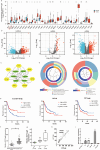
Pseudogene OCT4-pg5 upregulates OCT4B expression to promote bladder cancer progression by competing with miR-145-5p
- PMID: 38842275
- PMCID: PMC11229759
- DOI: 10.1080/15384101.2024.2353554
Pseudogene OCT4-pg5 upregulates OCT4B expression to promote bladder cancer progression by competing with miR-145-5p
Abstract
Bladder cancer (BC) is one of the most common malignant neoplasms worldwide. Competing endogenous RNA (ceRNA) networks may identify potential biomarkers associated with the progression and prognosis of BC. The OCT4-pg5/miR-145-5p/OCT4B ceRNA network was found to be related to the progression and prognosis of BC. OCT4-pg5 expression was significantly higher in BC cell lines than in normal bladder cells, with OCT4-pg5 expression correlating with OCT4B expression and advanced tumor grade. Overexpression of OCT4-pg5 and OCT4B promoted the proliferation and invasion of BC cells, whereas miR-145-5p suppressed these activities. The 3' untranslated region (3'UTR) of OCT4-pg5 competed for miR-145-5p, thereby increasing OCT4B expression. In addition, OCT4-pg5 promoted epithelial-mesenchymal transition (EMT) by activating the Wnt/β-catenin pathway and upregulating the expression of matrix metalloproteinases (MMPs) 2 and 9 as well as the transcription factors zinc finger E-box binding homeobox (ZEB) 1 and 2. Elevated expression of OCT4-pg5 and OCT4B reduced the sensitivity of BC cells to cisplatin by reducing apoptosis and increasing the proportion of cells in G1. The OCT4-pg5/miR-145-5p/OCT4B axis promotes the progression of BC by inducing EMT via the Wnt/β-catenin pathway and enhances cisplatin resistance. This axis may represent a therapeutic target in patients with BC.
Keywords: OCT4-pg5; OCT4B; bladder cancer; competing endogenous RNA; miR-145-5p.
Conflict of interest statement
No potential conflict of interest was reported by the author(s).
Figures


Similar articles
-
CEP55 3'-UTR promotes epithelial-mesenchymal transition and enhances tumorigenicity of bladder cancer cells by acting as a ceRNA regulating miR-497-5p.Cell Oncol (Dordr). 2022 Dec;45(6):1217-1236. doi: 10.1007/s13402-022-00712-6. Epub 2022 Nov 14. Cell Oncol (Dordr). 2022. PMID: 36374443
-
OCT4 pseudogene 5 upregulates OCT4 expression to promote proliferation by competing with miR-145 in endometrial carcinoma.Oncol Rep. 2015 Apr;33(4):1745-52. doi: 10.3892/or.2015.3763. Epub 2015 Jan 29. Oncol Rep. 2015. PMID: 25634023
-
Gambogic Acid Improves Cisplatin Resistance of Bladder Cancer Cells through the Epithelial-Mesenchymal Transition Pathway Mediated by the miR-205-5p/ZEB1 Axis.Ann Clin Lab Sci. 2024 May;54(3):354-362. Ann Clin Lab Sci. 2024. PMID: 39048172
-
LncRNA PVT1 accelerates malignant phenotypes of bladder cancer cells by modulating miR-194-5p/BCLAF1 axis as a ceRNA.Aging (Albany NY). 2020 Nov 16;12(21):22291-22312. doi: 10.18632/aging.202203. Epub 2020 Nov 16. Aging (Albany NY). 2020. PMID: 33188158 Free PMC article.
-
Long noncoding RNA H19 acts as a miR-340-3p sponge to promote epithelial-mesenchymal transition by regulating YWHAZ expression in paclitaxel-resistant breast cancer cells.Environ Toxicol. 2020 Sep;35(9):1015-1028. doi: 10.1002/tox.22938. Epub 2020 May 18. Environ Toxicol. 2020. PMID: 32420678
Cited by
-
PGK1 can affect the prognosis and development of bladder cancer.Cancer Med. 2024 Sep;13(18):e70242. doi: 10.1002/cam4.70242. Cancer Med. 2024. PMID: 39315723 Free PMC article.
-
MiR-145-5p inhibits proliferation of hepatocellular carcinoma, acting through PAI-1.Am J Transl Res. 2025 Feb 15;17(2):888-896. doi: 10.62347/NPLT8946. eCollection 2025. Am J Transl Res. 2025. PMID: 40092137 Free PMC article.
-
MicroRNA-145 in urologic tumors: biological roles, regulatory networks, and clinical translation.Front Pharmacol. 2025 Jul 4;16:1609646. doi: 10.3389/fphar.2025.1609646. eCollection 2025. Front Pharmacol. 2025. PMID: 40689207 Free PMC article. Review.
-
Non-coding RNAs in bladder cancer, a bridge between gut microbiota and host?Front Immunol. 2024 Nov 19;15:1482765. doi: 10.3389/fimmu.2024.1482765. eCollection 2024. Front Immunol. 2024. PMID: 39628486 Free PMC article. Review.
References
-
- Global Burden of Disease 2019 Cancer Collaboration, Kocarnik JM, Compton K, Dean FE, Fu W, Gaw BL, et al. Cancer incidence, mortality, years of life lost, years lived with disability, and disability-adjusted life years for 29 cancer groups from 2010 to 2019: a systematic analysis for the global burden of disease study 2019. JAMA Oncol. 2022;8(3):420–444. doi: 10.1001/jamaoncol.2021.6987 - DOI - PMC - PubMed
MeSH terms
Substances
LinkOut - more resources
Full Text Sources
Other Literature Sources
Medical
Research Materials
