. 2024 Aug 1;12(8):1022-1038.
doi: 10.1158/2326-6066.CIR-23-0636.

CLN-617 Retains IL2 and IL12 in Injected Tumors to Drive Robust and Systemic Immune-Mediated Antitumor Activity

Affiliations

CLN-617 Retains IL2 and IL12 in Injected Tumors to Drive Robust and Systemic Immune-Mediated Antitumor Activity

Naveen K Mehta et al. Cancer Immunol Res. .

Abstract

Despite clinical evidence of antitumor activity, the development of cytokine therapies has been hampered by a narrow therapeutic window and limited response rates. Two cytokines of high interest for clinical development are interleukin 2 (IL2) and interleukin 12 (IL12), which potently synergize to promote the activation and proliferation of T cells and NK cells. However, the only approved human IL2 therapy, Proleukin, is rarely used in the clinic due to systemic toxicities, and no IL12 product has been approved to date due to severe dose-limiting toxicities. Here, we describe CLN-617, a first-in-class therapeutic for intratumoral (IT) injection that co-delivers IL2 and IL12 on a single molecule in a safe and effective manner. CLN-617 is a single-chain fusion protein comprised of IL2, leukocyte-associated immunoglobulin-like receptor 2 (LAIR2), human serum albumin (HSA), and IL12. LAIR2 and HSA function to retain CLN-617 in the treated tumor by binding collagen and increasing molecular weight, respectively. We found that IT administration of a murine surrogate of CLN-617, mCLN-617, eradicated established treated and untreated tumors in syngeneic models, significantly improved response to anti-PD1 checkpoint therapy, and generated a robust abscopal response dependent on cellular immunity and antigen cross-presentation. CLN-617 is being evaluated in a clinical trial in patients with advanced solid tumors (NCT06035744).


Conflict of interest statement

N.K. Mehta reports personal fees from Cullinan Therapeutics outside the submitted work and has a pending patent for CLN-617. K. Rakhra reports other support from Cullinan Therapeutics outside the submitted work and has a pending patent for 63/579429 and for 63/593615. N. Momin reports a patent for US20230174623A1 licensed to Cullinan Therapeutics. K.D. Wittrup reports personal fees from Cullinan Therapeutics outside the submitted work and reports a patent for US20230174623A1 licenced to Cullinan Therapeutics. P.A. Baeuerle reports other support from Cullinan Oncology during the conduct of the study and personal fees and other support from Cullinan Therapeutics outside the submitted work. J.S. Michaelson reports personal fees from Cullinan Therapeutics outside the submitted work and has a pending patent for “Bi-functional linear fusion collagen-localized immunomodulatory molecules and methods thereof.” K. Rakhra, K.A. Meetze, P.A. Baeuerle, and J.S. Michaelson are employees and stock owners of Cullinan Therapeutics. N.K. Mehta and K.D. Wittrup own stock in Cullinan Therapeutics. Cullinan Therapeutics is the sponsor of an ongoing Phase I clinical trial for CLN-617. N. Momin and K.D. Wittrup report a patent US20230174623A1 licensed to Cullinan Therapeutics. N.K. Mehta, K. Rakhra, K.A. Meetze, P.A. Baeuerle and J.S. Michaelson are authors on pending patents 61/579429 and 63/593615 related to this work. No disclosures were reported by the other authors.

Figures
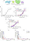
Figure 1.
Figure 1.
Bioactivity and in vivo pharmacokinetics of mCLN-617. A, Schematic of mCLN-617 design. B, ELISA-based binding assay on rat collagen I-coated plates, representative of three independent experiments. C, CTLL-2 or (D) 2D6 proliferation assay with normal or collagen-coated tissue culture plates, representative of four and two independent experiments, respectively. E,F, 50 pmol mCLN-617 was intratumorally injected in MC38-bearing mice and IL2 and IL12 levels detected in tumor and in serum at the indicated timepoints using MSD analysis.
Figure 2.
Figure 2.
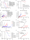
In vivo bioactivity of mCLN-617 in the injected tumor. A and B, Mice bearing ∼100 mm3 B16F10 tumors were intratumorally treated with the indicated test articles on days 0 and 6 (n = 10 mice/group). Shown are (A) survival curves and (B) body weight changes. C, Mice bearing B16F10 tumors were intratumorally or intravenously treated with the indicated dose levels of PBS or mCLN-617 on days 0, 6, and 12, and tumor growth kinetics are shown (n = 10 mice/group). D, Mice bearing ∼100 mm3 MC38 tumors were intratumorally treated with the indicated dose of mCLN-617 or PBS on days 0 and 6 or IP-treated with 10 mg/kg anti-PD1 antibody or isotype control BIW. Survival curves are shown (n = 10 mice/group). E, Mice were treated as in D but with 400 pmol mCLN-617 on the indicated days, and survival curves are shown (n = 10 mice/group). F, Mice were treated as in D but with 400 pmol mCLN-617 (n = 10 mice/group). Shown are body weight changes. G and H, mCLN-617-treated survivors from the 400 pmol group in D and age-matched naïve controls were rechallenged with MC38 tumors on the contralateral flank and Pan02 tumors on the ipsilateral flank. Shown are growth curves for MC38 (G) and Pan02 (H; n = 10 mice/group). In A, statistical significance was evaluated using the Mantel–Cox log-rank test. n.s. indicates that group comparisons LAIR1-MSA-IL2 + IL12-MSA-LAIR1 (10 pmol each) vs. CLN-617 (10 pmpl) and LAIR1-MSA-IL2 + IL12-MSA-LAIR1 (100 pmol each) vs CLN-617 (100 pmol) were not statistically significant. In B, body weight differences were compared using two-way ANOVA with multiple comparison’s test at each timepoint. For both A and B, LAIR1-MSA-2 (10 pmol) + 12-MSA-LAIR1 (10 pmol) was compared against mCLN-617 (10 pmol), and LAIR1-MSA-2 (100 pmol) + 12-MSA-LAIR1 (100 pmol) was compared against mCLN-617 (100 pmol).
Figure 3.
Figure 3.
Abscopal effect mediated by mCLN-617. A, Schematic of the dual flank MC38 tumor model used in B and C. B, Mice were intratumorally treated in their right MC38 flank tumor with 400 pmol mCLN-617 or PBS on days 0 and 5 with or without intraperitoneal treatment with 10 mg/kg anti-PD1 antibody BIW (n = 7–10 mice/group, and results are representative of two independent experiments). C, Mice were intratumorally treated in their right MC38 flank tumor with 50 (low), 100 (mid), or 200 pmol (high) mCLN-617 on days 0 and 5 and peripheral blood evaluated by flow cytometry on day 6. D, Schematic of MC38 liver metastasis model used in E. E, Mice were implanted with MC38 flank tumors and intrasplenically injected with MC38-luc cells, which seeded to the liver after resection of the spleen (n = 10 mice/group). The flank tumor was intratumorally treated with 200 pmol mCLN-617 or PBS on days 0 and 5 with or without intraperitoneal treatment with 10 mg/kg anti-PD1 antibody BIW. Shown are curves tracking flank tumor volume and luciferase signal. In C, statistical significance was evaluated with one-way ANOVA and Sidak’s multiple comparisons test.
Figure 4.
Figure 4.
Mechanism of action of mCLN-617. A, Mice bearing MC38 tumors were intratumorally treated with 1,000 pmol mCLN-617 or PBS and cytokine levels in the tumor measured by MSD 24 h after treatment (n = 5 mice/group). B–F, Mice were implanted with MC38 tumors in both flanks and intratumorally treated in their right MC38 flank tumor with 50, 100, or 200 pmol mCLN-617 on days 0 and 5 and treated and untreated tumors (B−D and F) and peripheral blood (E) evaluated by flow cytometry on day 6. G, Mice bearing two MC38 tumors were intratumorally treated in the right flank with 200 pmol mCLN-617 or PBS on days 0 and 6 (n = 10 mice/group). Approximately 24 h before first IT treatment, mice were IP administered 10 mg/kg depleting antibodies, and depletion continued BIW through the course of the study. Shown are tumor growth curves. H, WT or Batf3−/− C57BL/6 mice bearing two MC38 tumors were intratumorally treated in the right flank with 200 pmol mCLN-617 or PBS on days 0 and 6 (n = 10 mice/group). Shown are tumor growth curves. In A–F, statistical significance was evaluated with one-way ANOVA and Sidak’s multiple comparisons test. In G and H, tumor volumes were compared using two-way ANOVA with multiple comparison’s test at each timepoint. For G, each depletion group was compared against mCLN-617 with no depletion, and for H, the treated Batf3−/− mouse group was compared against the treated WT mouse group.
Figure 5.
Figure 5.
mCLN-617-mediated clonal expansion of tumor-associated T-cell clonotypes. Mice were implanted with MC38 tumors in both flanks and treated in the right flank intratumorally with mCLN-617 and IP with isotype control (Group 1; G1), intratumorally with mCLN-617 and IP with anti-PD1 (Group 2; G2), intratumorally with vehicle and IP with anti-PD1 (Group 3; G3), or intratumorally with vehicle and IP with isotype control (Group 4; G4; n = 4–5 mice/group). IT treatments were performed on days 0 and 5 and intraperitoneal treatments on days 0 and 3. mCLN-617 was administered at 200 pmol and antibodies at 10 mg/kg. Peripheral was collected on days 0 and 6, and tumors were collected on day 6 for TCRB sequencing (n = 4–5 per group). A, Simpson clonality was calculated for each group on day 0 (predose) and day 6 (postdose). B, Individual clonotypes were correlated between predose and postdose peripheral blood samples. Shown are scatterplots for individual representative mice, and each point represents the frequencies of a unique clonotype. Clonotypes are annotated based on association with the treated tumor. C, The Morisita index was calculated as a measure of correlation between predose and postdose peripheral blood samples. D, Newly expanded clones in each group were characterized as previously detected or newly detected postdose. E, Shown are the number of newly expanded clones in each mouse per group that is right (treated) or left (untreated) tumor associated.
Figure 6.
Figure 6.
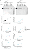
Characterization of CLN-617. A, CLN-617 was incubated in the indicated matrix for the indicated duration at 37°C and a Western blot performed detecting IL2 or IL12. B–I, Human PBMCs were CD3 stimulated overnight and then treated with the indicated test articles for 10 min (C and D), 2 h (B), 48 h (E–G and I), or 96 h (H). PBMC lysate was analyzed by signaling microarray (B) or CD8+ T cells assessed by flow cytometry (C–I). Data from one of the three representative donors is shown. In B, correlation analysis was assessed using Pearson correlation. Results from C–I are representative of two independent experiments.

References

    1. Spolski R, Li P, Leonard WJ. Biology and regulation of IL-2: from molecular mechanisms to human therapy. Nat Rev Immunol 2018;18:648–59. - PubMed
    1. Trinchieri G. Interleukin-12 and the regulation of innate resistance and adaptive immunity. Nat Rev Immunol 2003;3:133–46. - PubMed
    1. Nguyen KG, Vrabel MR, Mantooth SM, Hopkins JJ, Wagner ES, Gabaldon TA, et al. . Localized interleukin-12 for cancer immunotherapy. Front Immunol 2020;11:575597. - PMC - PubMed
    1. Bacon CM, McVicar DW, Ortaldo JR, Rees RC, O’Shea JJ, Johnston JA. Interleukin 12 (IL-12) induces tyrosine phosphorylation of JAK2 and TYK2: differential use of Janus family tyrosine kinases by IL-2 and IL-12. J Exp Med 1995;181:399–404. - PMC - PubMed
    1. Boyman O, Sprent J. The role of interleukin-2 during homeostasis and activation of the immune system. Nat Rev Immunol 2012;12:180–90. - PubMed

Associated data

LinkOut - more resources