. 2024 Sep 3;12(9):1170-1183.
doi: 10.1158/2326-6066.CIR-23-0527.

Pancreatic Epithelial IL17/IL17RA Signaling Drives B7-H4 Expression to Promote Tumorigenesis

Affiliations

Pancreatic Epithelial IL17/IL17RA Signaling Drives B7-H4 Expression to Promote Tumorigenesis

Susana Castro-Pando et al. Cancer Immunol Res. .

Abstract

IL17 is required for the initiation and progression of pancreatic cancer, particularly in the context of inflammation, as previously shown by genetic and pharmacological approaches. However, the cellular compartment and downstream molecular mediators of IL17-mediated pancreatic tumorigenesis have not been fully identified. This study examined the cellular compartment required by generating transgenic animals with IL17 receptor A (IL17RA), which was genetically deleted from either the pancreatic epithelial compartment or the hematopoietic compartment via generation of IL17RA-deficient (IL17-RA-/-) bone marrow chimeras, in the context of embryonically activated or inducible Kras. Deletion of IL17RA from the pancreatic epithelial compartment, but not from hematopoietic compartment, resulted in delayed initiation and progression of premalignant lesions and increased infiltration of CD8+ cytotoxic T cells to the tumor microenvironment. Absence of IL17RA in the pancreatic compartment affected transcriptional profiles of epithelial cells, modulating stemness, and immunological pathways. B7-H4, a known inhibitor of T-cell activation encoded by the gene Vtcn1, was the checkpoint molecule most upregulated via IL17 early during pancreatic tumorigenesis, and its genetic deletion delayed the development of pancreatic premalignant lesions and reduced immunosuppression. Thus, our data reveal that pancreatic epithelial IL17RA promotes pancreatic tumorigenesis by reprogramming the immune pancreatic landscape, which is partially orchestrated by regulation of B7-H4. Our findings provide the foundation of the mechanisms triggered by IL17 to mediate pancreatic tumorigenesis and reveal the avenues for early pancreatic cancer immune interception. See related Spotlight by Lee and Pasca di Magliano, p. 1130.


Conflict of interest statement

J.K. Kolls reports grants from NHLBI and grants from NIAID during the conduct of the study. J.P. Allison reports other support from Achelois, other support from Adaptive Biotechnologies, other support from Apricity, other support from BioAlta, other support from BioNTech, other support from Candel Therapeutics, other support from Dragonfly, other support from Earli, other support from Enable Medicine, other support from Hummingbird, other support from ImaginAb, other support from Lava Therapeutics, other support from Lytix, other support from Marker, other support from PBM Capital, other support from Phenomic AI, other support from Polaris Pharma, other support from Time Bioventures, other support from Trained Therapeutix, other support from Two Bear Capital, and other support from Venn Biosciences during the conduct of the study; other support from Achelois, other support from Adaptive Biotechnologies, other support from Apricity, other support from BioAtla, other support from BioNTech, other support from Candel Therapeutics, other support from Dragonfly, other support from Earli, other support from Enable Medcine, other support from Hummingbird, other support from ImaginAb, other support from Lava Therapeutics, other support from Lytix, other support from Marker, other support from PBM Capital, other support from Phenomic AI, other support from Polaris Pharma, other support from Time Bioventures, other support from Trained Therapeutix, other support from Two Bear Captial, and other support from Venn Biosciences outside the submitted work. G. Lozano reports grants from The University of Texas MDACC during the conduct of the study. F. McAllister reports personal fees from Neologics Bio outside the submitted work. No disclosures were reported by the other authors.

Figures
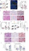
Figure 1.
Figure 1.
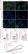
Epithelial pancreatic compartment is required for IL17/IL17RA signaling to induce tumorigenesis. A, Protocol for the collection of pancreata from KC and KC; IL17-RAfl/fl at 30 weeks of age. B, Representative immunofluorescence staining (10×) of DAPI (blue), E-cadherin (green), and IL17R (red) in pancreatic tissue of KC and KC; IL17RAfl/fl at 20 weeks of age. C, Representative staining (10×) on pancreatic tissue sections of KC and KC; IL17RAfl/fl at 30 weeks of hematoxylin and eosin (left), Alcian Blue staining (middle), and trichrome staining (right). D, Quantification of fractional cross-sectional area occupied by normal tissue and fibro-inflammatory infiltrate from KC and KC; IL17-RAfl/fl at 30 weeks of age. Two-tailed Student t-tests were used to compare the statistical differences between KC and KC; IL17-RAfl/fl mice at the same time point for each kind of lesion. E, Quantification of fractional cross-sectional area occupied by ADM, early PanIN and Advanced (Adv.) PanIN from KC and KC; IL17-RAfl/fl at 30 weeks of age. Two-tailed Student t-tests were used to compare the statistical differences between KC and KC; IL17-RAfl/fl mice at the same time point for each kind of lesion. F, Protocol for the collection/transplantation of the BM in KC mice at 20 or 30 weeks of age. G,Il17ra expression by qPCR of sorted CD45+ cells of KC mice transplanted with IL17RA+/+ or IL17RA−/− BM. H, Representative staining on pancreatic tissue sections of KC+IL17-RA+/+ BM and KC+IL17-RA−/− BM at 30 weeks of age. hematoxylin and eosin (left), Alcian Blue (medium), and trichrome staining (right). I, Quantification of fractional cross-sectional area occupied by normal tissue and fibro-inflammatory infiltrate from KC+IL17-RA+/+ BM and KC+IL17-RA−/− BM at 20 and 30 weeks of age. Two-tailed Student t-tests were used to compare the statistical differences between KC+IL17-RA+/+ BM and KC+IL17-RA−/− BM at the same time point for each kind of lesion, not statistically significant (ns). J, Quantification of fractional cross-sectional area occupied by ADM, early PanIN and Advanced (Adv.) PanIN from KC+IL17-RA+/+ BM and KC+IL17-RA−/− BM at 20 and 30 weeks of age. Two-tailed Student t-tests were used to compare the statistical differences between KC+IL17-RA+/+ BM and KC+IL17-RA−/− BM at the same time point for each kind of lesion, not statistically significant (ns).
Figure 2.
Figure 2.
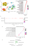
IL17/IL17RA pancreatic epithelial signaling modulates the tumor microenvironment. A, Representative Multi-IHC picture (10×) of pancreatic tissue KC and KC; IL17-RAfl/fl at 30 weeks of age. B–D, Quantification on pancreatic tissue sections of KC (black) and KC; IL17-RAfl/fl (red) at 30 weeks by Multiplex IHC of CD45+Ki67+ cells (B), CD45+CD3+CD8+ cells (C) and CD45+CD3+CD8+Granzyme B+ (Gzmb; D) cells. The statistical differences between groups were determined by two-tailed Student t-tests. E, Spatial analysis by L-function of CD8+ Gzmb+ cells.
Figure 3.
Figure 3.
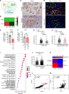
Loss of IL17RA promotes transcriptional changes in pancreatic epithelial cells. A, UMAP of scRNA-seq analysis from eight mice (4/group) of KC and KC; IL17-RAfl/fl at 30 weeks of age. B, GO analysis of all epithelial clusters. C, Comparison of PDAC associated Ras signature genes between epithelial clusters 2 and 3. D, GSEA analysis of the Epithelial Cluster 3. E, Radar chart showing how stem cell types and Epithelial Cluster 3 overlap. The values plotted represent the significance of the overlap (with each radar representing −log10P value).
Figure 4.
Figure 4.
IL17/IL17RA pancreatic epithelial signaling regulates the expression of B7-H4. A, Gene database comparisons with genes from Epithelial Cluster 3. B, Heatmap of immune checkpoint protein expression in the pancreas of KC and KC; IL17-RAfl/fl at 30 weeks as measured by qPCR. C, Representative picture of B7-H4 IHC (10×) of KC mice at 20 weeks (top) and 30 weeks of age (bottom). D, Representative picture of DAPI (blue), E-cadherin (green), and B7-H4 (red) IF staining on pancreatic tissue sections (10×) of KC (top) and KC; IL17-RAfl/fl at 30 weeks of age (bottom). E, Quantification of B7-H4 IF staining on pancreatic tissue sections of KC and KC; IL17-RAfl/fl mice at 30 Weeks of age. F, Quantification of B7-H4 staining on pancreatic tissue sections (10×) KC mice following BM transplantation from IL17RA+/+ or IL17RA−/− mice. G, Violin Plot of Eomes expression in CD8+ T-cells cluster. H, Violin Plot of Eomes expression in Exhausted CD8+ T-cells cluster. I, GO Analysis of CD8 T-cells cluster. J, Violin Plot of Vtcn1 expression in epithelial cluster from single-cell data from Carpenter and colleagues. K, Heatmap of Vtcn expression in epithelial cluster and Eomes expression in CD8+ T cells cluster from adjacent normal and PDAC patient’s pancreas tissue from single-cell data from Steele and colleagues. L, Correlation analysis of Eomes and Cd8a expression in 170 patients with PDAC from the Cancer Genome Atlas. M, Correlation analysis of Eomes and Il17ra expression in 170 patients with PDAC from the Cancer Genome Atlas.
Figure 5.
Figure 5.
B7-H4 promotes pancreatic tumorigenesis. A, Protocol for the collection of pancreata from KC and KC; B7-H4−/− at 30 weeks of age. B, Representative staining (10×) on pancreatic tissue sections of KC and KC; IL17RAfl/fl at 30 weeks of age for hematoxylin and eosin (left), Alcian Blue (middle) and trichrome staining (right). C–E, Quantification of Advanced PanINs (C), Early PanINs (D) and ADMs (E) in KC and KC; B7-H4−/− mice at 30 weeks of age. F, Representative picture of CD8 IHC staining (10×) on pancreatic tissue from KC (top) and KC; B7-H4−/− mice (bottom) at 30 weeks of age. G, Quantification of CD8+ cells. H, Representative picture of Gzmb staining on pancreatic tissue KC (top) and KC; B7-H4−/− mice (bottom) at 30 weeks. I, Quantification of Granzyme B (Gzmb)+ cells.

References

    1. Rahib L, Wehner MR, Matrisian LM, Nead KT. Estimated projection of US cancer incidence and death to 2040. JAMA Netw Open 2021;4:e214708. - PMC - PubMed
    1. Rhim AD, Mirek ET, Aiello NM, Maitra A, Bailey JM, McAllister F, et al. EMT and dissemination precede pancreatic tumor formation. Cell 2012;148:349–61. - PMC - PubMed
    1. Ho WJ, Jaffee EM, Zheng L. The tumour microenvironment in pancreatic cancer - clinical challenges and opportunities. Nat Rev Clin Oncol 2020;17:527–40. - PMC - PubMed
    1. Ren B, Cui M, Yang G, Wang H, Feng M, You L, et al. Tumor microenvironment participates in metastasis of pancreatic cancer. Mol Cancer 2018;17:108. - PMC - PubMed
    1. Provenzano PP, Cuevas C, Chang AE, Goel VK, Von Hoff DD, Hingorani SR. Enzymatic targeting of the stroma ablates physical barriers to treatment of pancreatic ductal adenocarcinoma. Cancer Cell 2012;21:418–29. - PMC - PubMed

MeSH terms

Substances